Department of Anatomy and Structural Biology, Graduate School of Medicine, University of Yamanashi, 1110 Shimokato, Chuo, Yamanashi, 409-3898, Japan.
Department of Cell Biology and Anatomy, Graduate School of Medicine, the University of Tokyo, 7-3-1 Hongo, Bunkyo-ku, Tokyo, 113-0033, Japan.
Commun Biol. 2020 Oct 16;3(1):585. doi: 10.1038/s42003-020-01321-5.
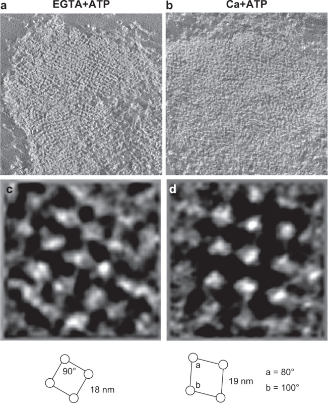
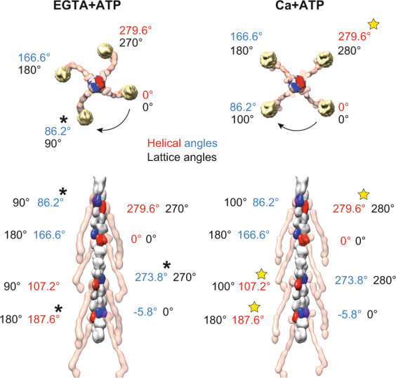
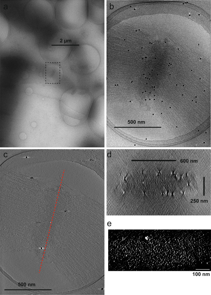
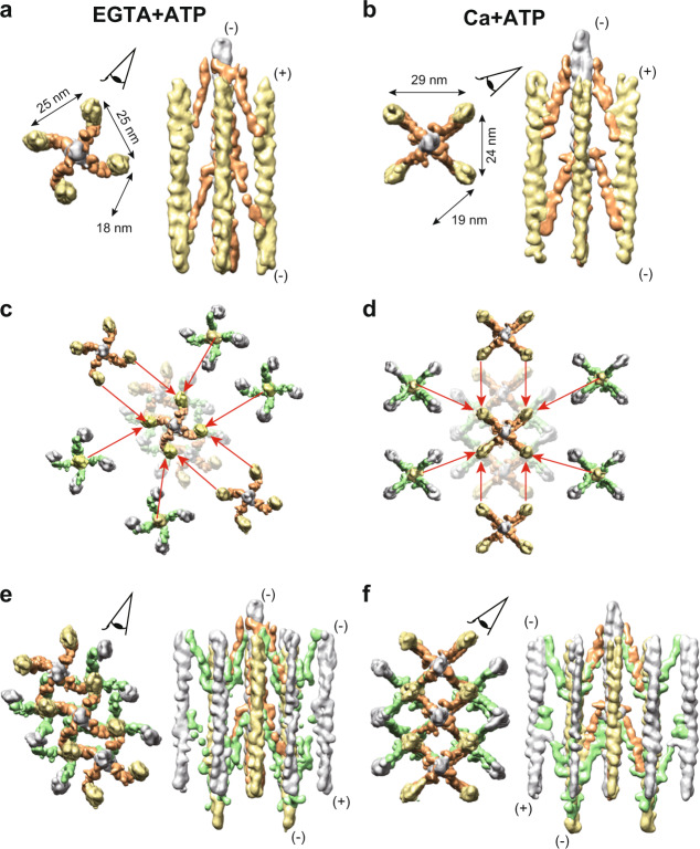
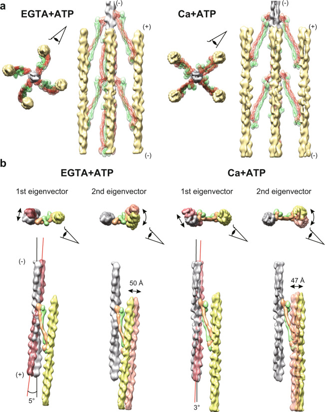
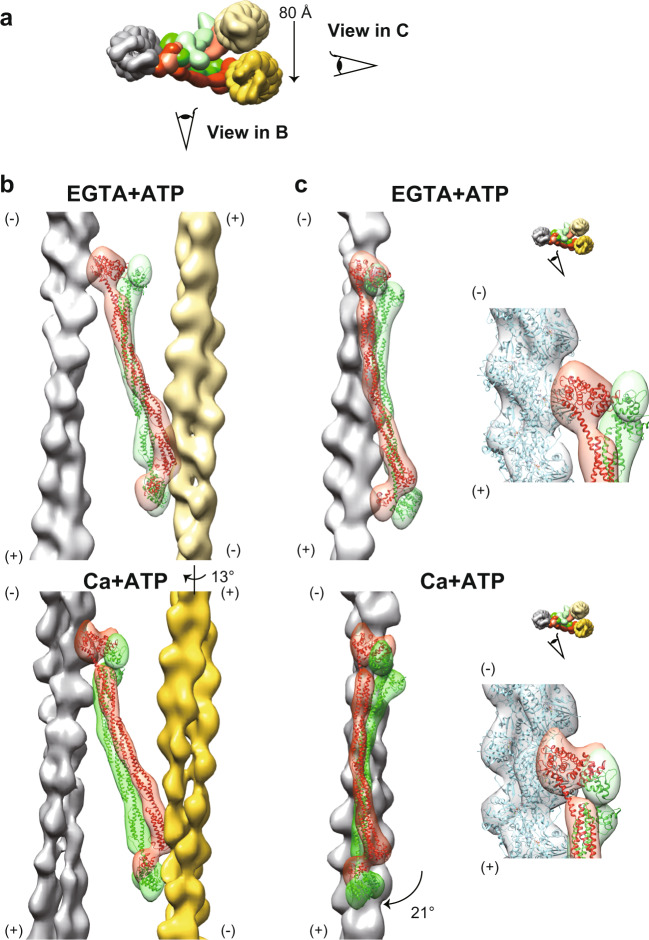
The Z-disc forms a boundary between sarcomeres, which constitute structural and functional units of striated muscle tissue. Actin filaments from adjacent sarcomeres are cross-bridged by α-actinin in the Z-disc, allowing transmission of tension across the myofibril. Despite decades of studies, the 3D structure of Z-disc has remained elusive due to the limited resolution of conventional electron microscopy. Here, we observed porcine cardiac myofibrils using cryo-electron tomography and reconstructed the 3D structures of the actin-actinin cross-bridging complexes within the Z-discs in relaxed and activated states. We found that the α-actinin dimers showed contraction-dependent swinging and sliding motions in response to a global twist in the F-actin lattice. Our observation suggests that the actin-actinin complex constitutes a molecular lattice spring, which maintains the integrity of the Z-disc during the muscle contraction cycle.
Z 盘形成了肌节的边界,肌节是横纹肌组织的结构和功能单位。来自相邻肌节的肌动蛋白丝通过 Z 盘中的α-辅肌动蛋白交联,从而使张力在肌原纤维上传递。尽管经过了几十年的研究,但由于传统电子显微镜的分辨率有限,Z 盘的 3D 结构仍然难以捉摸。在这里,我们使用冷冻电子断层扫描观察了猪心肌纤维,并重建了松弛和激活状态下 Z 盘中肌动蛋白-辅肌动蛋白交联复合物的 3D 结构。我们发现,α-辅肌动蛋白二聚体在 F-肌动蛋白晶格的整体扭曲下表现出收缩依赖性的摆动和滑动运动。我们的观察表明,肌动蛋白-辅肌动蛋白复合物构成了分子晶格弹簧,在肌肉收缩循环中保持 Z 盘的完整性。