• 文献检索
  • 文档翻译
  • 深度研究
  • 学术资讯
  • Suppr Zotero 插件Zotero 插件
  • 邀请有礼
  • 套餐&价格
  • 历史记录
应用&插件
Suppr Zotero 插件Zotero 插件浏览器插件Mac 客户端Windows 客户端微信小程序
定价
高级版会员购买积分包购买API积分包
服务
文献检索文档翻译深度研究API 文档MCP 服务
关于我们
关于 Suppr公司介绍联系我们用户协议隐私条款
关注我们

Suppr 超能文献

核心技术专利:CN118964589B侵权必究
粤ICP备2023148730 号-1Suppr @ 2026

文献检索

告别复杂PubMed语法,用中文像聊天一样搜索,搜遍4000万医学文献。AI智能推荐,让科研检索更轻松。

立即免费搜索

文件翻译

保留排版,准确专业,支持PDF/Word/PPT等文件格式,支持 12+语言互译。

免费翻译文档

深度研究

AI帮你快速写综述,25分钟生成高质量综述,智能提取关键信息,辅助科研写作。

立即免费体验

建立并验证新生儿大脑功能成像头模型数据库。

Construction and validation of a database of head models for functional imaging of the neonatal brain.

机构信息

DOT-HUB, Department of Medical Physics and Biomedical Engineering, University College London, London, UK.

Biomedical Optics Research Laboratory, Medical Physics and Biomedical Engineering, University College London, London, UK.

出版信息

Hum Brain Mapp. 2021 Feb 15;42(3):567-586. doi: 10.1002/hbm.25242. Epub 2020 Oct 17.

DOI:10.1002/hbm.25242
PMID:33068482
原文链接:https://pmc.ncbi.nlm.nih.gov/articles/PMC7814762/
Abstract

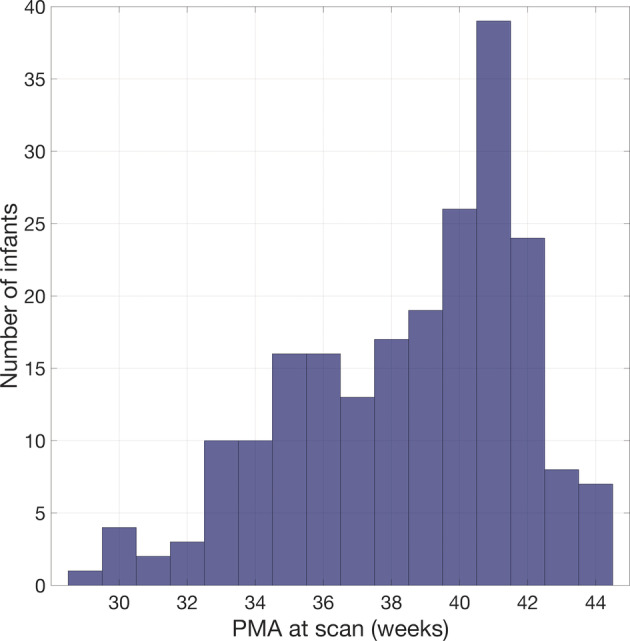
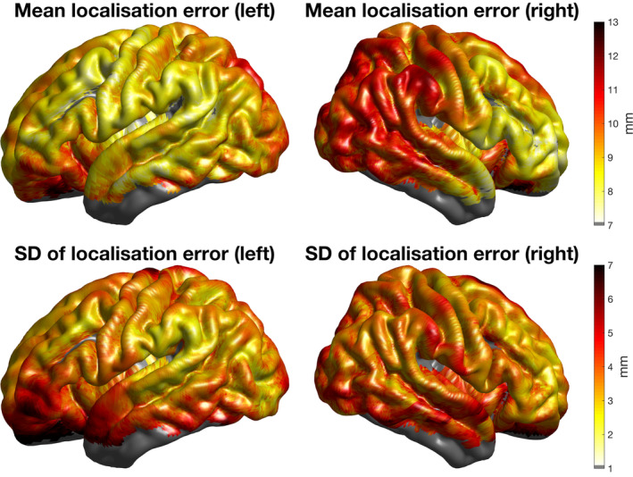
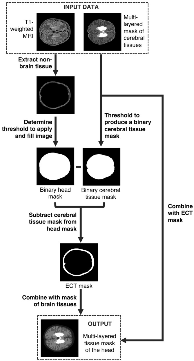
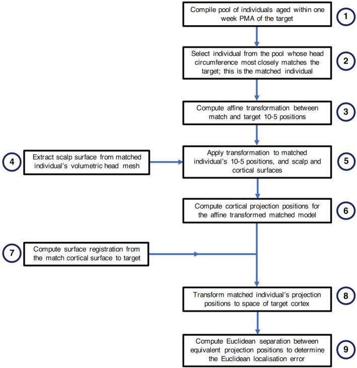
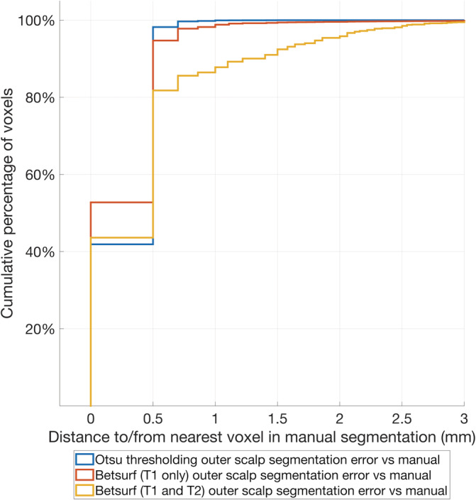
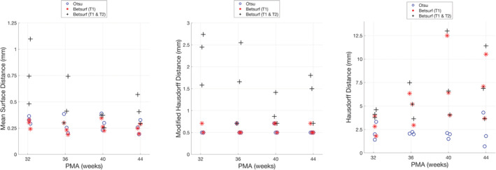
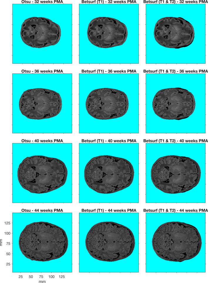
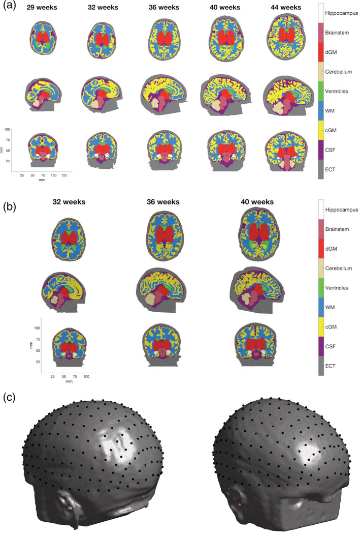
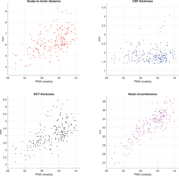
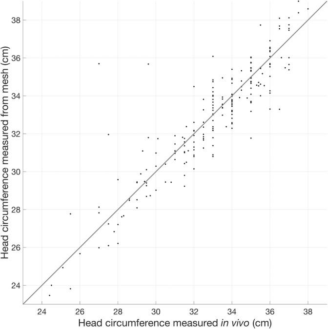
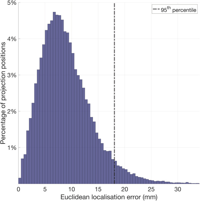
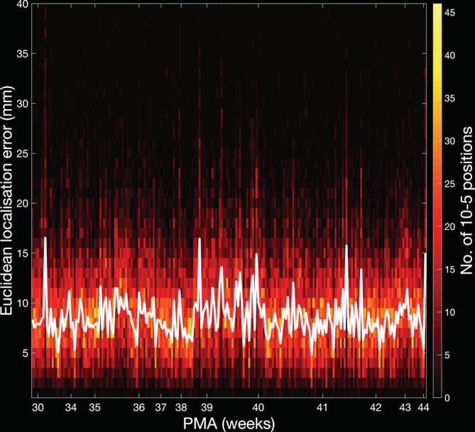
The neonatal brain undergoes dramatic structural and functional changes over the last trimester of gestation. The accuracy of source localisation of brain activity recorded from the scalp therefore relies on accurate age-specific head models. Although an age-appropriate population-level atlas could be used, detail is lost in the construction of such atlases, in particular with regard to the smoothing of the cortical surface, and so such a model is not representative of anatomy at an individual level. In this work, we describe the construction of a database of individual structural priors of the neonatal head using 215 individual-level datasets at ages 29-44 weeks postmenstrual age from the Developing Human Connectome Project. We have validated a method to segment the extra-cerebral tissue against manual segmentation. We have also conducted a leave-one-out analysis to quantify the expected spatial error incurred with regard to localising functional activation when using a best-matching individual from the database in place of a subject-specific model; the median error was calculated to be 8.3 mm (median absolute deviation 3.8 mm). The database can be applied for any functional neuroimaging modality which requires structural data whereby the physical parameters associated with that modality vary with tissue type and is freely available at www.ucl.ac.uk/dot-hub.

摘要

胎儿大脑在妊娠最后三个月经历显著的结构和功能变化。因此,头皮记录的脑活动源定位的准确性依赖于准确的年龄特异性头部模型。尽管可以使用适合年龄的人群水平图谱,但在构建此类图谱时会丢失细节,特别是在皮质表面的平滑化方面,因此此类模型不能代表个体水平的解剖结构。在这项工作中,我们描述了使用来自发育人类连接组计划的 215 个个体水平数据集,在妊娠后 29-44 周的年龄范围内,构建个体新生儿头部结构先验数据库的方法。我们已经验证了一种针对大脑外组织进行分割的方法,以对抗手动分割。我们还进行了一次留一分析,以量化在使用数据库中最佳匹配的个体代替特定于个体的模型来定位功能激活时所涉及的预期空间误差;中位数误差计算为 8.3 毫米(中位数绝对偏差为 3.8 毫米)。该数据库可应用于任何需要结构数据的功能神经影像学模式,其中与该模式相关的物理参数随组织类型而变化,并可在 www.ucl.ac.uk/dot-hub 上免费获得。

https://cdn.ncbi.nlm.nih.gov/pmc/blobs/29b0/7814762/62495cf082de/HBM-42-567-g012.jpg
https://cdn.ncbi.nlm.nih.gov/pmc/blobs/29b0/7814762/68df63496298/HBM-42-567-g001.jpg
https://cdn.ncbi.nlm.nih.gov/pmc/blobs/29b0/7814762/40d145b77ea6/HBM-42-567-g002.jpg
https://cdn.ncbi.nlm.nih.gov/pmc/blobs/29b0/7814762/91342dc1c9f8/HBM-42-567-g003.jpg
https://cdn.ncbi.nlm.nih.gov/pmc/blobs/29b0/7814762/4da800d8ef08/HBM-42-567-g004.jpg
https://cdn.ncbi.nlm.nih.gov/pmc/blobs/29b0/7814762/e57f4bde37f4/HBM-42-567-g005.jpg
https://cdn.ncbi.nlm.nih.gov/pmc/blobs/29b0/7814762/228cdc508d38/HBM-42-567-g006.jpg
https://cdn.ncbi.nlm.nih.gov/pmc/blobs/29b0/7814762/afbb9cf974bc/HBM-42-567-g007.jpg
https://cdn.ncbi.nlm.nih.gov/pmc/blobs/29b0/7814762/71622d6fdd9c/HBM-42-567-g008.jpg
https://cdn.ncbi.nlm.nih.gov/pmc/blobs/29b0/7814762/73bb88a9617d/HBM-42-567-g009.jpg
https://cdn.ncbi.nlm.nih.gov/pmc/blobs/29b0/7814762/e6c7da55466d/HBM-42-567-g010.jpg
https://cdn.ncbi.nlm.nih.gov/pmc/blobs/29b0/7814762/db84e5dfa3f6/HBM-42-567-g011.jpg
https://cdn.ncbi.nlm.nih.gov/pmc/blobs/29b0/7814762/62495cf082de/HBM-42-567-g012.jpg

相似文献

1
Construction and validation of a database of head models for functional imaging of the neonatal brain.建立并验证新生儿大脑功能成像头模型数据库。
Hum Brain Mapp. 2021 Feb 15;42(3):567-586. doi: 10.1002/hbm.25242. Epub 2020 Oct 17.
2
A 4D neonatal head model for diffuse optical imaging of pre-term to term infants.一种用于早产儿至足月儿漫射光学成像的四维新生儿头部模型。
Neuroimage. 2014 Oct 15;100:385-94. doi: 10.1016/j.neuroimage.2014.06.028. Epub 2014 Jun 18.
3
Detail-preserving construction of neonatal brain atlases in space-frequency domain.在空间频率域中进行保留细节的新生儿脑图谱构建。
Hum Brain Mapp. 2016 Jun;37(6):2133-50. doi: 10.1002/hbm.23160. Epub 2016 Mar 14.
4
Effects of atlas-based anatomy on modelled light transport in the neonatal head.基于寰枢椎解剖结构的模型对新生儿头部光传输的影响。
Phys Med Biol. 2023 Jul 3;68(13):135019. doi: 10.1088/1361-6560/acd48c.
5
Patch-based augmentation of Expectation-Maximization for brain MRI tissue segmentation at arbitrary age after premature birth.基于补丁增强的期望最大化算法用于早产后脑MRI组织分割(适用于任意年龄)
Neuroimage. 2016 Feb 15;127:387-408. doi: 10.1016/j.neuroimage.2015.12.009. Epub 2015 Dec 17.
6
Construction of 4D high-definition cortical surface atlases of infants: Methods and applications.婴儿4D高清皮质表面图谱的构建:方法与应用
Med Image Anal. 2015 Oct;25(1):22-36. doi: 10.1016/j.media.2015.04.005. Epub 2015 Apr 17.
7
Brain structure-function associations identified in large-scale neuroimaging data.在大规模神经影像数据中识别出的脑结构-功能关联。
Brain Struct Funct. 2016 Dec;221(9):4459-4474. doi: 10.1007/s00429-015-1177-6. Epub 2016 Jan 9.
8
A 4D infant brain volumetric atlas based on the UNC/UMN baby connectome project (BCP) cohort.基于 UNC/UMN 婴儿连接组计划 (BCP) 队列的 4D 婴儿脑容量图谱。
Neuroimage. 2022 Jun;253:119097. doi: 10.1016/j.neuroimage.2022.119097. Epub 2022 Mar 14.
9
Construction of multi-region-multi-reference atlases for neonatal brain MRI segmentation.构建新生儿脑 MRI 分割的多区域-多参考图谱。
Neuroimage. 2010 Jun;51(2):684-93. doi: 10.1016/j.neuroimage.2010.02.025. Epub 2010 Feb 17.
10
Age-specific gray and white matter DTI atlas for human brain at 33, 36 and 39 postmenstrual weeks.33、36 和 39 孕周胎儿大脑特定年龄的灰白质弥散张量成像图谱。
Neuroimage. 2019 Jan 15;185:685-698. doi: 10.1016/j.neuroimage.2018.06.069. Epub 2018 Jun 26.

引用本文的文献

1
Robust photogrammetric scalp morphology estimation for functional optical neuroimaging.用于功能性光学神经成像的稳健摄影测量头皮形态估计
Neurophotonics. 2025 Jul;12(3):035002. doi: 10.1117/1.NPh.12.3.035002. Epub 2025 Jul 28.
2
Effects of atlas-based anatomy on modelled light transport in the neonatal head.基于寰枢椎解剖结构的模型对新生儿头部光传输的影响。
Phys Med Biol. 2023 Jul 3;68(13):135019. doi: 10.1088/1361-6560/acd48c.
3
The Developing Human Connectome Project Neonatal Data Release.人类连接组计划新生儿数据发布

本文引用的文献

1
Cerebral hemodynamic responses in preterm-born neonates to visual stimulation: classification according to subgroups and analysis of frontotemporal-occipital functional connectivity.早产新生儿对视觉刺激的脑血流动力学反应:根据亚组分类及额颞枕功能连接分析
Neurophotonics. 2019 Oct;6(4):045005. doi: 10.1117/1.NPh.6.4.045005. Epub 2019 Nov 6.
2
Changes of functional response in sensorimotor cortex of preterm and full-term infants during the first year: An fNIRS study.早产儿和足月儿大脑感觉运动皮层功能反应变化:一项近红外光谱研究。
Early Hum Dev. 2019 Jun;133:23-28. doi: 10.1016/j.earlhumdev.2019.04.007. Epub 2019 Apr 29.
3
Front Neurosci. 2022 May 23;16:886772. doi: 10.3389/fnins.2022.886772. eCollection 2022.
4
The neural correlates of inhibitory control in 10-month-old infants: A functional near-infrared spectroscopy study.10 个月大婴儿抑制控制的神经相关性:一项功能近红外光谱研究。
Neuroimage. 2022 Aug 15;257:119241. doi: 10.1016/j.neuroimage.2022.119241. Epub 2022 May 7.
5
Group-level cortical functional connectivity patterns using fNIRS: assessing the effect of bilingualism in young infants.使用功能近红外光谱技术(fNIRS)的组水平皮质功能连接模式:评估双语对幼儿的影响。
Neurophotonics. 2021 Apr;8(2):025011. doi: 10.1117/1.NPh.8.2.025011. Epub 2021 Jun 12.
6
Longitudinal infant fNIRS channel-space analyses are robust to variability parameters at the group-level: An image reconstruction investigation.纵向婴儿近红外光谱通道空间分析对组水平的变异性参数具有稳健性:图像重建研究。
Neuroimage. 2021 Aug 15;237:118068. doi: 10.1016/j.neuroimage.2021.118068. Epub 2021 Apr 26.
Transcranial focused ultrasound neuromodulation of the human primary motor cortex.
经颅聚焦超声人脑初级运动皮层神经调控。
Sci Rep. 2018 Jul 3;8(1):10007. doi: 10.1038/s41598-018-28320-1.
4
Somatotopic Mapping of the Developing Sensorimotor Cortex in the Preterm Human Brain.发育中早产儿大脑体感运动皮层的躯体定位图。
Cereb Cortex. 2018 Jul 1;28(7):2507-2515. doi: 10.1093/cercor/bhy050.
5
Construction of a neonatal cortical surface atlas using Multimodal Surface Matching in the Developing Human Connectome Project.利用发展中的人类连接组计划中的多模态表面匹配构建新生儿皮质表面图谱。
Neuroimage. 2018 Oct 1;179:11-29. doi: 10.1016/j.neuroimage.2018.06.018. Epub 2018 Jun 14.
6
The developing human connectome project: A minimal processing pipeline for neonatal cortical surface reconstruction.人类连接组计划:新生儿皮质表面重建的最小处理流程。
Neuroimage. 2018 Jun;173:88-112. doi: 10.1016/j.neuroimage.2018.01.054. Epub 2018 Jan 31.
7
Multimodal surface matching with higher-order smoothness constraints.多模态曲面匹配的高阶平滑约束。
Neuroimage. 2018 Feb 15;167:453-465. doi: 10.1016/j.neuroimage.2017.10.037. Epub 2017 Oct 31.
8
Computationally Efficient Algorithms for Sparse, Dynamic Solutions to the EEG Source Localization Problem.计算高效的 EEG 源定位问题稀疏动态解算法。
IEEE Trans Biomed Eng. 2018 Jun;65(6):1359-1372. doi: 10.1109/TBME.2017.2739824. Epub 2017 Sep 14.
9
Numerical evaluation of the skull for human neuromodulation with transcranial focused ultrasound.颅部数值评估用于经颅聚焦超声的人类神经调节。
J Neural Eng. 2017 Dec;14(6):066012. doi: 10.1088/1741-2552/aa843e.
10
Macroanatomical Landmarks Featuring Junctions of Major Sulci and Fissures and Scalp Landmarks Based on the International 10-10 System for Analyzing Lateral Cortical Development of Infants.基于国际10-10系统的婴儿外侧皮质发育分析中,以主要脑沟和脑裂交界处为特征的大体解剖标志及头皮标志。
Front Neurosci. 2017 Jul 11;11:394. doi: 10.3389/fnins.2017.00394. eCollection 2017.