Marino Natascia, German Rana, Rao Xi, Simpson Ed, Liu Sheng, Wan Jun, Liu Yunlong, Sandusky George, Jacobsen Max, Stoval Miranda, Cao Sha, Storniolo Anna Maria V
Susan G. Komen Tissue Bank at the IU Simon Cancer Center, Indianapolis, IN 46202 USA.
Department of Medicine, Indiana University School of Medicine, Indianapolis, IN 46202 USA.
NPJ Breast Cancer. 2020 Oct 6;6:50. doi: 10.1038/s41523-020-00191-8. eCollection 2020.
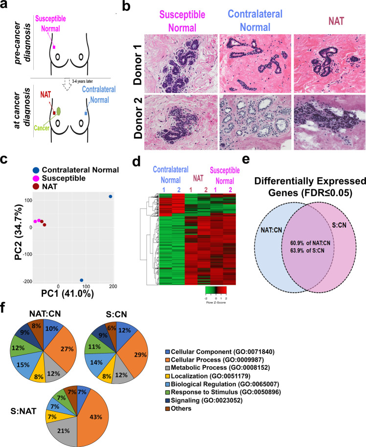
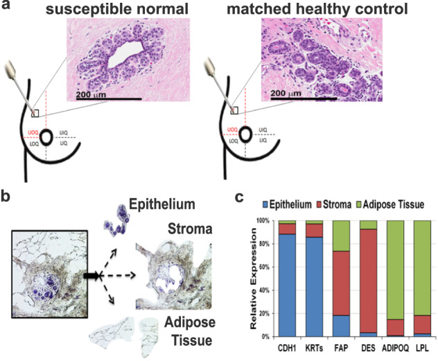
Histologically normal tissue adjacent to the tumor can provide insight of the microenvironmental alterations surrounding the cancerous lesion and affecting the progression of the disease. However, little is known about the molecular changes governing cancer initiation in cancer-free breast tissue. Here, we employed laser microdissection and whole-transcriptome profiling of the breast epithelium prior to and post tumor diagnosis to identify the earliest alterations in breast carcinogenesis. Furthermore, a comprehensive analysis of the three tissue compartments (microdissected epithelium, stroma, and adipose tissue) was performed on the breast donated by either healthy subjects or women prior to the clinical manifestation of cancer (labeled "susceptible normal tissue"). Although both susceptible and healthy breast tissues appeared histologically normal, the susceptible breast epithelium displayed a significant upregulation of genes involved in fatty acid uptake/transport (CD36 and AQP7), lipolysis (LIPE), and lipid peroxidation (AKR1C1). Upregulation of lipid metabolism- and fatty acid transport-related genes was observed also in the microdissected susceptible stromal and adipose tissue compartments, respectively, when compared with the matched healthy controls. Moreover, inter-compartmental co-expression analysis showed increased epithelium-adipose tissue crosstalk in the susceptible breasts as compared with healthy controls. Interestingly, reductions in natural killer (NK)-related gene signature and CD45+/CD20+ cell staining were also observed in the stromal compartment of susceptible breasts. Our study yields new insights into the cancer initiation process in the breast. The data suggest that in the early phase of cancer development, metabolic activation of the breast, together with increased epithelium-adipose tissue crosstalk may create a favorable environment for final cell transformation, proliferation, and survival.
肿瘤旁组织学正常的组织能够为了解癌性病变周围的微环境改变以及影响疾病进展的因素提供线索。然而,对于无癌乳腺组织中控制癌症起始的分子变化却知之甚少。在此,我们利用激光显微切割技术以及肿瘤诊断前后乳腺上皮细胞的全转录组分析,来确定乳腺癌发生过程中最早出现的变化。此外,我们还对健康受试者或癌症临床表现出现之前的女性捐赠的乳腺组织(标记为“易感正常组织”)的三个组织部分(显微切割的上皮组织、基质组织和脂肪组织)进行了全面分析。尽管易感乳腺组织和健康乳腺组织在组织学上均表现正常,但易感乳腺上皮细胞中参与脂肪酸摄取/转运(CD36和AQP7)、脂肪分解(LIPE)以及脂质过氧化(AKR1C1)的基因显著上调。与匹配的健康对照相比,在显微切割的易感基质组织和脂肪组织部分中也分别观察到脂质代谢和脂肪酸转运相关基因的上调。此外,与健康对照相比,易感乳腺组织中上皮组织与脂肪组织之间的跨室共表达分析显示二者之间的相互作用增强。有趣的是,在易感乳腺组织的基质部分还观察到自然杀伤(NK)相关基因特征以及CD45+/CD20+细胞染色减少。我们的研究为乳腺癌症起始过程提供了新的见解。数据表明,在癌症发展的早期阶段,乳腺的代谢激活以及上皮组织与脂肪组织之间相互作用的增强可能为最终的细胞转化、增殖和存活创造有利环境。