Department of Medicine, University of Washington, Seattle, Washington State, USA.
Department of Psychiatry and Behavioral Sciences, Division of Child and Adolescent Psychiatry, Johns Hopkins University School of Medicine, Baltimore, Maryland, USA.
Pediatr Obes. 2021 Apr;16(4):e12732. doi: 10.1111/ijpo.12732. Epub 2020 Oct 20.
Quantitative magnetic resonance imaging (MRI) evidence of mediobasal hypothalamic (MBH) gliosis positively correlates with body mass index (BMI) in adults. This has neither been well explored in children nor have other brain regions involved in appetitive processing been tested for evidence of gliosis.
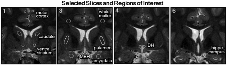
Multi-site cross-sectional study in children to test for differences in quantitative T2 signal (measure of gliosis) by region and to assess relationships with age and BMI. Participants underwent brain MRI using the same equipment and protocol to quantify T2 relaxation time in six bilateral regions of interest (ROIs): putamen, caudate, ventral striatum, amygdala, hippocampus and MBH, and three control regions: white matter, motor cortex and dorsal hypothalamus.
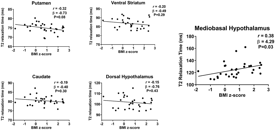
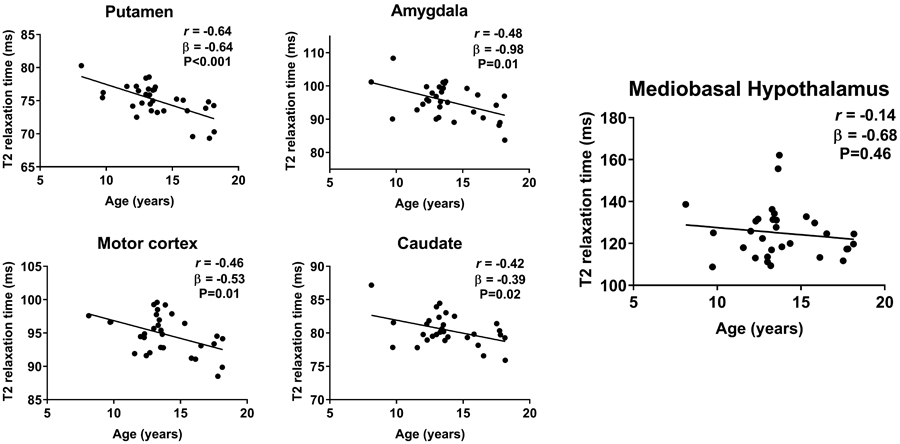
Thirty-one participants (61% female) were included in a combined sample from the University of Washington (N = 9) and John Hopkins University (N = 22). Mean age was 14 ± 3 years, and BMI z-score was 0.7 ± 1.1 (26% with obesity). No study site-related differences were seen in T2 relaxation time across all nine regions (chi (8): 9.46, P = .30). Regional differences in T2 relaxation time were present (P < .001). MBH presented longer T2 relaxation time, suggestive of gliosis, when compared to all regions (P < .001), including an intra-hypothalamic control. Physiological age-related declines in T2 relaxation times were found in grey matter ROIs, but not in the MBH (r = -0.14, P = .46). MBH was the only region with a positive correlation between T2 relaxation time and BMI z-score (r = 0.38, P = .03).
In a multi-site study, pilot data suggest that quantitative MRI detected normal maturation-related brain variation as well as evidence that MBH gliosis is associated with increased adiposity in children.
定量磁共振成像(MRI)显示中脑下丘脑(MBH)神经胶质增生与成人的体重指数(BMI)呈正相关。这在儿童中尚未得到充分研究,也没有对参与食欲处理的其他脑区进行神经胶质增生的证据检测。
对儿童进行多中心横断面研究,以测试不同脑区定量 T2 信号(神经胶质增生的测量指标)的差异,并评估其与年龄和 BMI 的关系。参与者使用相同的设备和方案进行脑 MRI,以量化六个双侧感兴趣区(ROI)的 T2 弛豫时间:壳核、尾状核、腹侧纹状体、杏仁核、海马体和 MBH,以及三个对照区:白质、运动皮层和背侧下丘脑。
来自华盛顿大学(N = 9)和约翰霍普金斯大学(N = 22)的 31 名参与者(61%为女性)被纳入合并样本。平均年龄为 14 ± 3 岁,BMI z 分数为 0.7 ± 1.1(26%有肥胖症)。在所有 9 个区域,T2 弛豫时间在研究地点之间没有差异(χ (8):9.46,P =.30)。T2 弛豫时间存在区域差异(P < .001)。与所有区域(P < .001)相比,包括下丘脑内对照区,MBH 的 T2 弛豫时间较长,提示有神经胶质增生。在灰质 ROI 中发现了与生理年龄相关的 T2 弛豫时间下降,但在 MBH 中没有(r = -0.14,P =.46)。MBH 是唯一与 BMI z 分数呈正相关的区域(r = 0.38,P =.03)。
在一项多中心研究中,初步数据表明,定量 MRI 检测到正常的与成熟相关的脑区变化,以及 MBH 神经胶质增生与儿童肥胖症有关的证据。