• 文献检索
  • 文档翻译
  • 深度研究
  • 学术资讯
  • Suppr Zotero 插件Zotero 插件
  • 邀请有礼
  • 套餐&价格
  • 历史记录
应用&插件
Suppr Zotero 插件Zotero 插件浏览器插件Mac 客户端Windows 客户端微信小程序
定价
高级版会员购买积分包购买API积分包
服务
文献检索文档翻译深度研究API 文档MCP 服务
关于我们
关于 Suppr公司介绍联系我们用户协议隐私条款
关注我们

Suppr 超能文献

核心技术专利:CN118964589B侵权必究
粤ICP备2023148730 号-1Suppr @ 2026

文献检索

告别复杂PubMed语法,用中文像聊天一样搜索,搜遍4000万医学文献。AI智能推荐,让科研检索更轻松。

立即免费搜索

文件翻译

保留排版,准确专业,支持PDF/Word/PPT等文件格式,支持 12+语言互译。

免费翻译文档

深度研究

AI帮你快速写综述,25分钟生成高质量综述,智能提取关键信息,辅助科研写作。

立即免费体验

衰老与鼻窦周围淋巴间隙体积呈正相关:使用3T黑血磁共振成像进行评估

Aging Is Positively Associated with Peri-Sinus Lymphatic Space Volume: Assessment Using 3T Black-Blood MRI.

作者信息

Park Mina, Kim Jin Woo, Ahn Sung Jun, Cha Yoon Jin, Suh Sang Hyun

机构信息

Department of Radiology, Gangnam Severance Hospital, Yonsei University College of Medicine, Seoul 06273, Korea.

Department of Pathology, Gangnam Severance Hospital, Yonsei University College of Medicine, Seoul 06273, Korea.

出版信息

J Clin Med. 2020 Oct 19;9(10):3353. doi: 10.3390/jcm9103353.

DOI:10.3390/jcm9103353
PMID:33086702
原文链接:https://pmc.ncbi.nlm.nih.gov/articles/PMC7590154/
Abstract

OBJECTIVES

Aging is a major risk factor for many neurological disorders and is associated with dural lymphatic dysfunction. We sought to evaluate the association of aging with the volume of the peri-sinus lymphatic space using contrast-enhanced 3T T1-weighted black-blood magnetic resonance imaging (MRI).

METHODS

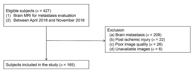
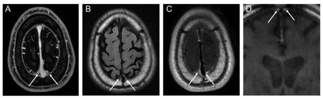
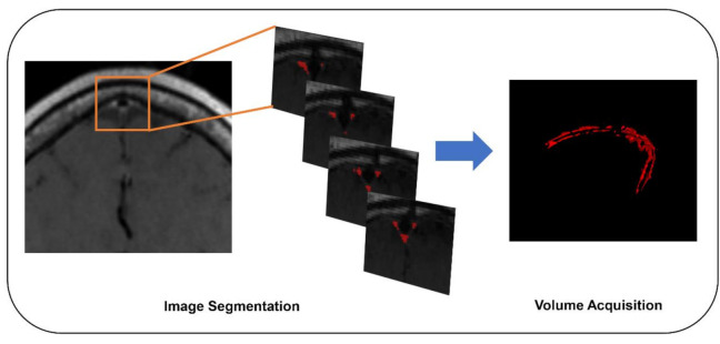
In this retrospective study, 165 presumed neurologically normal subjects underwent brain MRIs for cancer staging between April and November 2018. The parasagittal peri-sinus lymphatic space was evaluated using contrast-enhanced 3D T1-weighted black-blood MRIs, and volumes were measured with semiautomatic method. We compared the volumes of normalized peri-sinus lymphatic spaces between the elderly (≥65 years, = 72) and non-elderly ( = 93) groups and performed multivariate logistic regression analyses to assess if aging is independently associated with the volume of normalized peri-sinus lymphatic spaces.

RESULTS

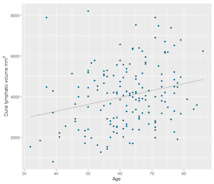
The normalized peri-sinus lymphatic space volume was significantly higher in the elderly than in the non-elderly (mean, 3323 ± 758.7 mL vs. 2968.7 ± 764.3 mL, = 0.047). After adjusting the intracranial volume, age age was the strongest factor independently associated with peri-sinus lymphatic space volume (β coefficient, 28.4 (5.7-51.2), = 0.015) followed by male sex (β coefficient, 672.4 (113.5-1230.8), = 0.019).

CONCLUSIONS

We found that the peri-sinus dural lymphatic space volume was higher in the elderly group than in the non-elderly group, and the increased peri-sinus lymphatic space was independently associated with aging. These findings indicate that the peri-sinus lymphatic space may be related with the aging process and lymphatic system dysfunction as well.

摘要

目的

衰老为许多神经疾病的主要风险因素,且与硬脑膜淋巴管功能障碍相关。我们试图通过对比增强3T T1加权黑血磁共振成像(MRI)评估衰老与窦周淋巴间隙容积之间的关联。

方法

在这项回顾性研究中,165名假定神经功能正常的受试者于2018年4月至11月间接受脑部MRI以进行癌症分期。使用对比增强3D T1加权黑血MRI评估矢状窦旁窦周淋巴间隙,并采用半自动方法测量其容积。我们比较了老年组(≥65岁,n = 72)和非老年组(n = 93)之间标准化窦周淋巴间隙的容积,并进行多因素逻辑回归分析,以评估衰老是否与标准化窦周淋巴间隙的容积独立相关。

结果

老年组标准化窦周淋巴间隙容积显著高于非老年组(均值分别为3323±758.7 mL和2968.7±764.3 mL,P = 0.047)。在校正颅内容积后,年龄是与窦周淋巴间隙容积独立相关的最强因素(β系数为28.4(5.7 - 51.2),P = 0.015),其次是男性(β系数为672.4(113.5 - 1230.8),P = 0.019)。

结论

我们发现老年组的窦周硬脑膜淋巴间隙容积高于非老年组,且窦周淋巴间隙增加与衰老独立相关。这些发现表明,窦周淋巴间隙可能与衰老过程以及淋巴系统功能障碍有关。

https://cdn.ncbi.nlm.nih.gov/pmc/blobs/2be1/7590154/73a189d3d414/jcm-09-03353-g004.jpg
https://cdn.ncbi.nlm.nih.gov/pmc/blobs/2be1/7590154/ed7e15373cf1/jcm-09-03353-g001.jpg
https://cdn.ncbi.nlm.nih.gov/pmc/blobs/2be1/7590154/3cc5f9a19b0b/jcm-09-03353-g002.jpg
https://cdn.ncbi.nlm.nih.gov/pmc/blobs/2be1/7590154/c354823b7d52/jcm-09-03353-g003.jpg
https://cdn.ncbi.nlm.nih.gov/pmc/blobs/2be1/7590154/73a189d3d414/jcm-09-03353-g004.jpg

相似文献

1
Aging Is Positively Associated with Peri-Sinus Lymphatic Space Volume: Assessment Using 3T Black-Blood MRI.衰老与鼻窦周围淋巴间隙体积呈正相关:使用3T黑血磁共振成像进行评估
J Clin Med. 2020 Oct 19;9(10):3353. doi: 10.3390/jcm9103353.
2
Assessment of Meningeal Lymphatics in the Parasagittal Dural Space: A Prospective Feasibility Study Using Dynamic Contrast-Enhanced Magnetic Resonance Imaging.评估矢状窦旁硬脑膜下腔脑膜淋巴管:使用动态对比增强磁共振成像的前瞻性可行性研究。
Korean J Radiol. 2023 May;24(5):444-453. doi: 10.3348/kjr.2022.0980. Epub 2023 Apr 12.
3
Performance of spin-echo and gradient-echo T1-weighted sequences for evaluation of dural venous sinus thrombosis and stenosis.自旋回波和梯度回波 T1 加权序列在硬脑膜静脉窦血栓和狭窄评估中的性能。
AJR Am J Roentgenol. 2013 Jul;201(1):162-9. doi: 10.2214/AJR.12.9095.
4
MRI diagnosis of dural sinus - Cortical venous thrombosis: Immediate post-contrast 3D GRE T1-weighted imaging versus unenhanced MR venography and conventional MR sequences.硬脑膜窦-皮质静脉血栓形成的MRI诊断:对比剂注射后即刻三维梯度回波T1加权成像与非增强磁共振静脉血管造影及传统磁共振序列的比较
Clin Neurol Neurosurg. 2015 Jul;134:44-54. doi: 10.1016/j.clineuro.2015.04.013. Epub 2015 Apr 22.
5
[Brain volumetric MRI study in healthy elderly persons using statistical parametric mapping].[使用统计参数映射对健康老年人进行脑容积磁共振成像研究]
Seishin Shinkeigaku Zasshi. 2004;106(2):138-51.
6
Impairment of the Glymphatic Pathway and Putative Meningeal Lymphatic Vessels in the Aging Human.衰老过程中神经胶质淋巴通路和推测性脑膜淋巴管的损伤。
Ann Neurol. 2020 Mar;87(3):357-369. doi: 10.1002/ana.25670. Epub 2020 Jan 30.
7
Diagnosis of non-acute cerebral venous thrombosis with 3D T1-weighted black blood sequence at 3T.3T磁共振3D T1加权黑血序列诊断非急性脑静脉血栓形成
J Neurol Sci. 2016 Aug 15;367:46-50. doi: 10.1016/j.jns.2016.05.052. Epub 2016 May 26.
8
Magnetic Resonance Imaging and Histopathological Visualization of Human Dural Lymphatic Vessels.人体硬脑膜淋巴管的磁共振成像与组织病理学可视化
Bio Protoc. 2018 Apr 20;8(8). doi: 10.21769/BioProtoc.2819.
9
Glymphatic MRI in idiopathic normal pressure hydrocephalus.特发性正常压力脑积水的脑淋巴系统磁共振成像
Brain. 2017 Oct 1;140(10):2691-2705. doi: 10.1093/brain/awx191.
10
Renal and renal sinus fat volumes as quantified by magnetic resonance imaging in subjects with prediabetes, diabetes, and normal glucose tolerance.磁共振成像定量分析糖尿病前期、糖尿病和正常葡萄糖耐量受试者的肾脏和肾窦脂肪体积。
PLoS One. 2020 Feb 19;15(2):e0216635. doi: 10.1371/journal.pone.0216635. eCollection 2020.

引用本文的文献

1
Morphologic and functional alterations in the parasagittal dural space in mild cognitive impairment.轻度认知障碍患者矢状窦旁硬脑膜间隙的形态学和功能改变
Sci Rep. 2025 Jul 4;15(1):23882. doi: 10.1038/s41598-025-07909-3.
2
Fluid and Waste Clearance in Central Nervous System Health and Diseases.中枢神经系统健康与疾病中的液体和废物清除
Neurodegener Dis. 2025 May 7:1-22. doi: 10.1159/000546018.
3
Vascular Contribution to Cerebral Waste Clearance Affected by Aging or Diabetes.衰老或糖尿病对脑血管清除脑内废物功能的影响

本文引用的文献

1
Cerebrospinal fluid tracer efflux to parasagittal dura in humans.脑脊髓液示踪剂向人脑矢状窦旁硬脑膜的外溢。
Nat Commun. 2020 Jan 17;11(1):354. doi: 10.1038/s41467-019-14195-x.
2
Impairment of the Glymphatic Pathway and Putative Meningeal Lymphatic Vessels in the Aging Human.衰老过程中神经胶质淋巴通路和推测性脑膜淋巴管的损伤。
Ann Neurol. 2020 Mar;87(3):357-369. doi: 10.1002/ana.25670. Epub 2020 Jan 30.
3
Contrast-enhanced modified 3D T1-weighted TSE black-blood imaging can improve detection of infectious and neoplastic meningitis.
Diagnostics (Basel). 2025 Apr 16;15(8):1019. doi: 10.3390/diagnostics15081019.
4
Long-term physical exercise facilitates putative glymphatic and meningeal lymphatic vessel flow in humans.长期体育锻炼有助于促进人体中假定的类淋巴系统和脑膜淋巴管的流动。
Nat Commun. 2025 Apr 9;16(1):3360. doi: 10.1038/s41467-025-58726-1.
5
Parasagittal dural volume correlates with cerebrospinal fluid volume and developmental delay in children with autism spectrum disorder.矢状旁硬脑膜容积与自闭症谱系障碍儿童的脑脊液容积及发育迟缓相关。
Commun Med (Lond). 2024 Oct 4;4(1):191. doi: 10.1038/s43856-024-00622-8.
6
Imaging evaluation and volumetric measurement of the space surrounding the diploic veins.硬脑膜窦周围间隙的影像学评估与容积测量。
Jpn J Radiol. 2024 Sep;42(9):953-961. doi: 10.1007/s11604-024-01572-w. Epub 2024 Apr 26.
7
Deep learning segmentation of peri-sinus structures from structural magnetic resonance imaging: validation and normative ranges across the adult lifespan.基于结构磁共振成像的鼻窦周围结构深度学习分割:成人全生命周期的验证和正常范围。
Fluids Barriers CNS. 2024 Feb 13;21(1):15. doi: 10.1186/s12987-024-00516-w.
8
Imaging of brain barrier inflammation and brain fluid drainage in human neurological diseases.脑屏障炎症和脑液引流的影像学在人类神经疾病中的应用。
Cell Mol Life Sci. 2024 Jan 12;81(1):31. doi: 10.1007/s00018-023-05073-3.
9
Non-invasive flow mapping of parasagittal meningeal lymphatics using 2D interslice flow saturation MRI.采用 2D 层间流饱和 MRI 无创性显示矢状窦旁脑膜淋巴管。
Fluids Barriers CNS. 2023 May 26;20(1):37. doi: 10.1186/s12987-023-00446-z.
10
Parasagittal dural space hypertrophy and amyloid-β deposition in Alzheimer's disease.阿尔茨海默病中矢状旁硬脑膜间隙肥大与β淀粉样蛋白沉积
Brain Commun. 2023 Apr 18;5(3):fcad128. doi: 10.1093/braincomms/fcad128. eCollection 2023.
对比增强改良 3D T1 加权 TSE 黑血成像可提高感染性和肿瘤性脑膜炎的检出率。
Eur Radiol. 2020 Feb;30(2):866-876. doi: 10.1007/s00330-019-06475-3. Epub 2019 Nov 5.
4
The Space between the Pial Sheath and the Cortical Venous Wall May Connect to the Meningeal Lymphatics.软脑膜鞘与皮质静脉壁之间的空间可能与脑膜淋巴管相通。
Magn Reson Med Sci. 2020 Feb 10;19(1):1-4. doi: 10.2463/mrms.bc.2019-0099. Epub 2019 Oct 15.
5
Meningeal lymphatic vessels at the skull base drain cerebrospinal fluid.颅底脑膜淋巴管引流脑脊液。
Nature. 2019 Aug;572(7767):62-66. doi: 10.1038/s41586-019-1419-5. Epub 2019 Jul 24.
6
Cortical cerebral blood flow in ageing: effects of haematocrit, sex, ethnicity and diabetes.大脑皮质脑血流的老化:血细胞比容、性别、种族和糖尿病的影响。
Eur Radiol. 2019 Oct;29(10):5549-5558. doi: 10.1007/s00330-019-06096-w. Epub 2019 Mar 18.
7
Myelin loss in white matter hyperintensities and normal-appearing white matter of cognitively impaired patients: a quantitative synthetic magnetic resonance imaging study.认知障碍患者的白质高信号区和正常外观白质中的髓鞘丢失:一项定量综合磁共振成像研究。
Eur Radiol. 2019 Sep;29(9):4914-4921. doi: 10.1007/s00330-018-5836-x. Epub 2018 Nov 28.
8
Meningeal Lymphatic Vessel Flow Runs Countercurrent to Venous Flow in the Superior Sagittal Sinus of the Human Brain.人脑上矢状窦中脑膜淋巴管的流动与静脉血流方向相反。
Tomography. 2018 Sep;4(3):99-104. doi: 10.18383/j.tom.2018.00013.
9
Significance of hyperintense arteries on Gd-enhanced 3D T1W black-blood imaging in acute stroke.钆增强 3D T1W 黑血成像上高信号动脉在急性脑卒中的意义。
Eur Radiol. 2019 Mar;29(3):1329-1337. doi: 10.1007/s00330-018-5669-7. Epub 2018 Aug 7.
10
Functional aspects of meningeal lymphatics in ageing and Alzheimer's disease.脑膜淋巴管在衰老和阿尔茨海默病中的功能方面。
Nature. 2018 Aug;560(7717):185-191. doi: 10.1038/s41586-018-0368-8. Epub 2018 Jul 25.