Sele Silvano, Liem Franziskus, Mérillat Susan, Jäncke Lutz
Division Neuropsychology, Department of Psychology, University of Zurich, Zurich, Switzerland.
University Research Priority Program (URPP), "Dynamics of Healthy Aging", University of Zurich, Zurich, Switzerland.
Front Hum Neurosci. 2020 Sep 4;14:363. doi: 10.3389/fnhum.2020.00363. eCollection 2020.
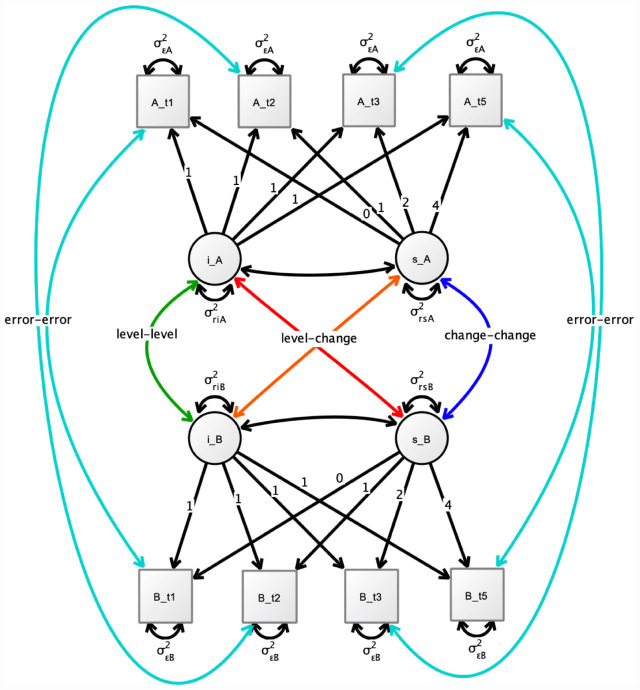
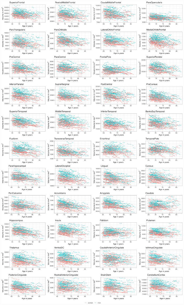
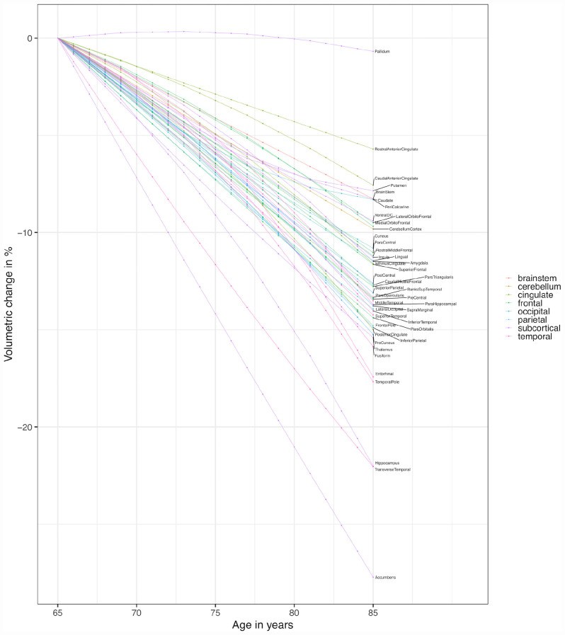
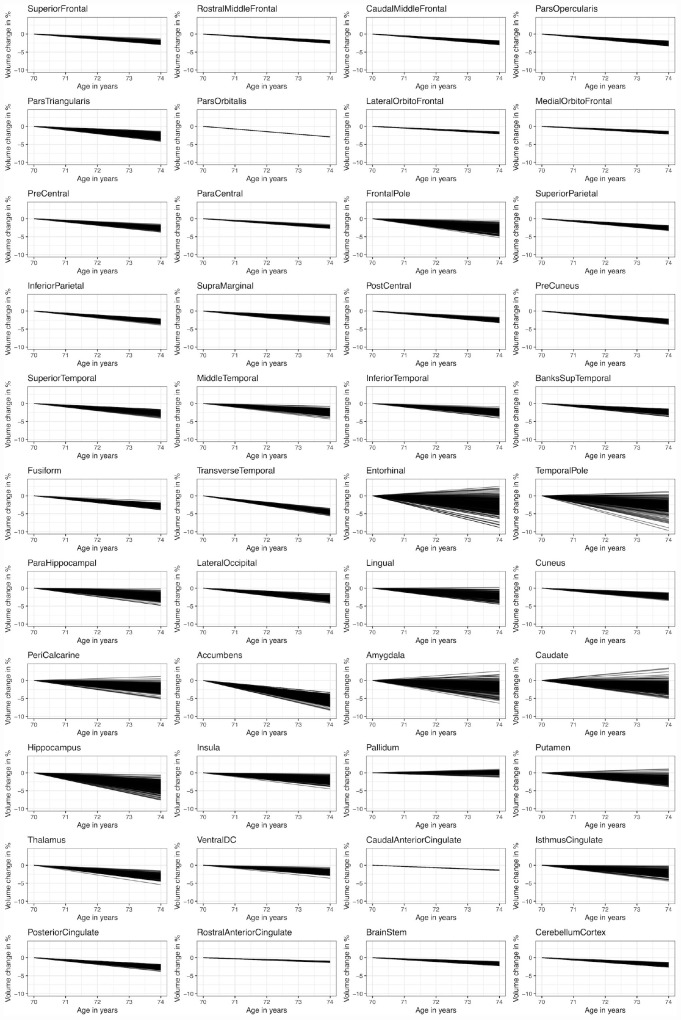
Describing the trajectories of age-related change for different brain structures has been of interest in many recent studies. However, our knowledge regarding these trajectories and their associations is still limited due to small sample sizes and low numbers of repeated measures. For the present study, we used a large longitudinal dataset (four measurements over 4 years) comprising anatomical data from a sample of healthy older adults ( = 231 at baseline). This dataset enables us to gain new insights about volumetric cortical and subcortical changes and their associations in the context of healthy aging. Brain structure volumes were derived from T1-weighted MRI scans using FreeSurfer segmentation tools. Brain structure trajectories were fitted using mixed models and latent growth curve models to gain information about the mean extent and variability of decline trajectories for different brain structures as well as the associations between individual trajectories. On the group level, our analyses indicate similar linear changes for frontal and parietal brain regions, while medial temporal regions showed an accelerated decline with advancing age. Regarding subcortical regions, some structures showed strong declines (e.g., hippocampus), others showed little decline (e.g., pallidum). Our data provide little evidence for sex differences regarding the aforementioned trajectories. Between-person variability of the person-specific slopes (random slopes) was largest in subcortical and medial temporal brain structures. When looking at the associations between the random slopes from each brain structure, we found that the decline is largely homogenous across the majority of cortical brain structures. In subcortical and medial temporal brain structures, however, more heterogeneity of the decline was observed, meaning that the extent of the decline in one structure is less predictive of the decline in another structure. Taken together, our study contributes to enhancing our understanding of structural brain aging by demonstrating (1) that average volumetric change differs across the brain and (2) that there are regional differences with respect to between-person variability in the slopes. Moreover, our data suggest (3) that random slopes are highly correlated across large parts of the cerebral cortex but (4) that some brain regions (i.e., medial temporal regions) deviate from this homogeneity.
描述不同脑结构与年龄相关的变化轨迹是最近许多研究的兴趣所在。然而,由于样本量小和重复测量次数少,我们对这些轨迹及其关联的了解仍然有限。在本研究中,我们使用了一个大型纵向数据集(4年内进行4次测量),该数据集包含来自健康老年人样本(基线时n = 231)的解剖学数据。这个数据集使我们能够在健康衰老的背景下,对皮质和皮质下体积变化及其关联有新的认识。脑结构体积是使用FreeSurfer分割工具从T1加权MRI扫描中得出的。使用混合模型和潜在生长曲线模型拟合脑结构轨迹,以获取不同脑结构衰退轨迹的平均程度和变异性以及个体轨迹之间关联的信息。在组水平上,我们的分析表明额叶和顶叶脑区有相似的线性变化,而内侧颞叶区域随着年龄增长衰退加速。关于皮质下区域,一些结构衰退明显(如海马体),另一些结构衰退很少(如苍白球)。我们的数据几乎没有提供关于上述轨迹存在性别差异的证据。个体特异性斜率(随机斜率)的个体间变异性在皮质下和内侧颞叶脑结构中最大。当观察每个脑结构的随机斜率之间的关联时,我们发现大多数皮质脑结构的衰退在很大程度上是同质的。然而,在皮质下和内侧颞叶脑结构中,观察到衰退的异质性更大,这意味着一个结构的衰退程度对另一个结构的衰退预测性较低。总之,我们的研究通过证明(1)大脑各区域的平均体积变化不同,以及(2)斜率的个体间变异性存在区域差异,有助于增进我们对脑结构衰老的理解。此外,我们的数据表明(3)随机斜率在大脑皮层的大部分区域高度相关,但(4)一些脑区(即内侧颞叶区域)偏离了这种同质性。