Pirkmajer Sergej, Bezjak Katja, Matkovič Urška, Dolinar Klemen, Jiang Lake Q, Miš Katarina, Gros Katarina, Milovanova Kseniya, Pirkmajer Katja Perdan, Marš Tomaž, Kapilevich Leonid, Chibalin Alexander V
Institute of Pathophysiology, Faculty of Medicine, University of Ljubljana, Ljubljana, Slovenia.
Integrative Physiology, Department of Molecular Medicine and Surgery, Karolinska Institutet, Stockholm, Sweden.
Front Physiol. 2020 Sep 25;11:566584. doi: 10.3389/fphys.2020.566584. eCollection 2020.
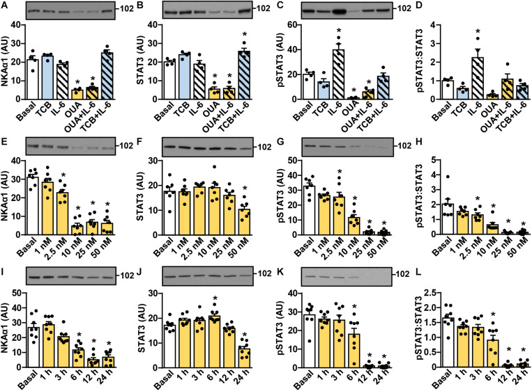
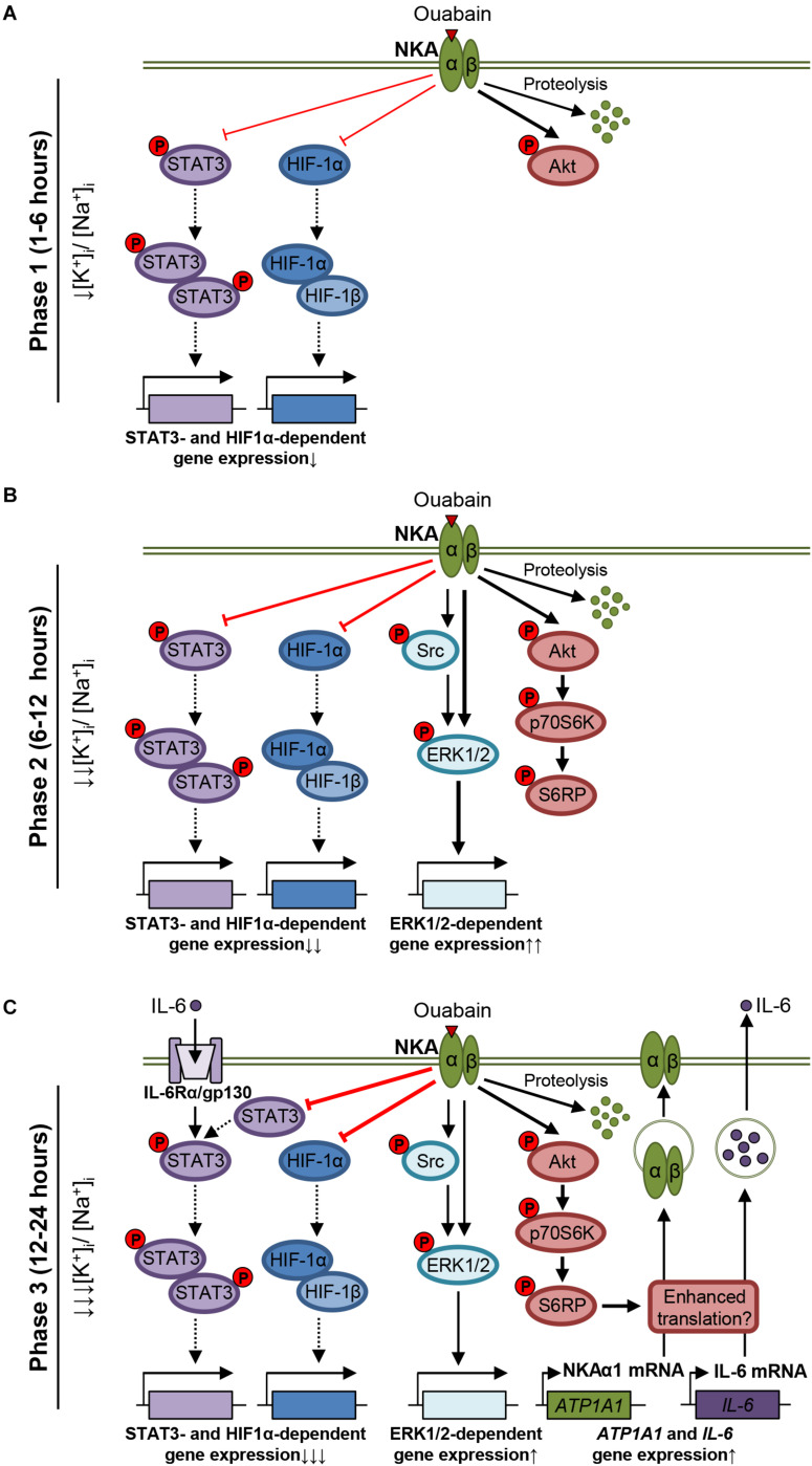
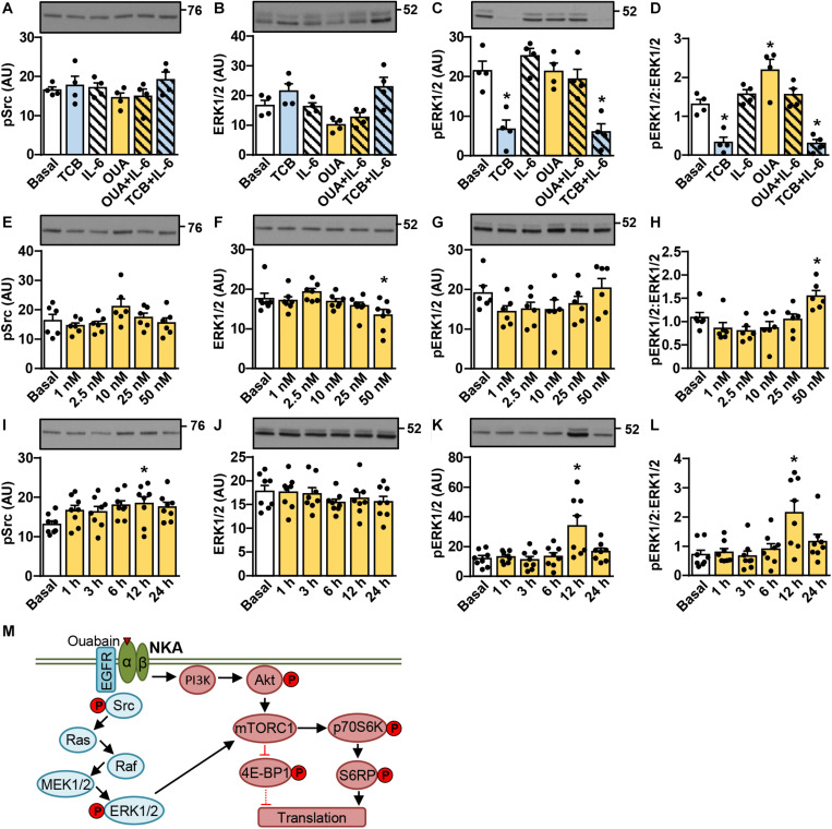
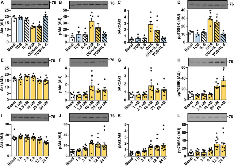
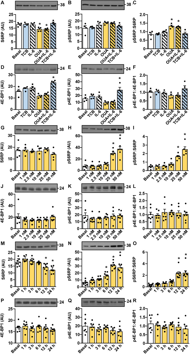
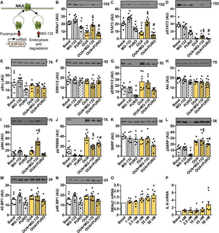
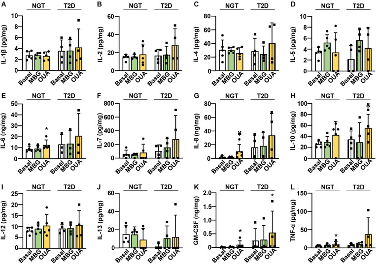
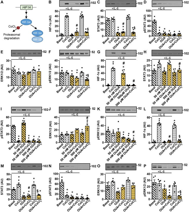
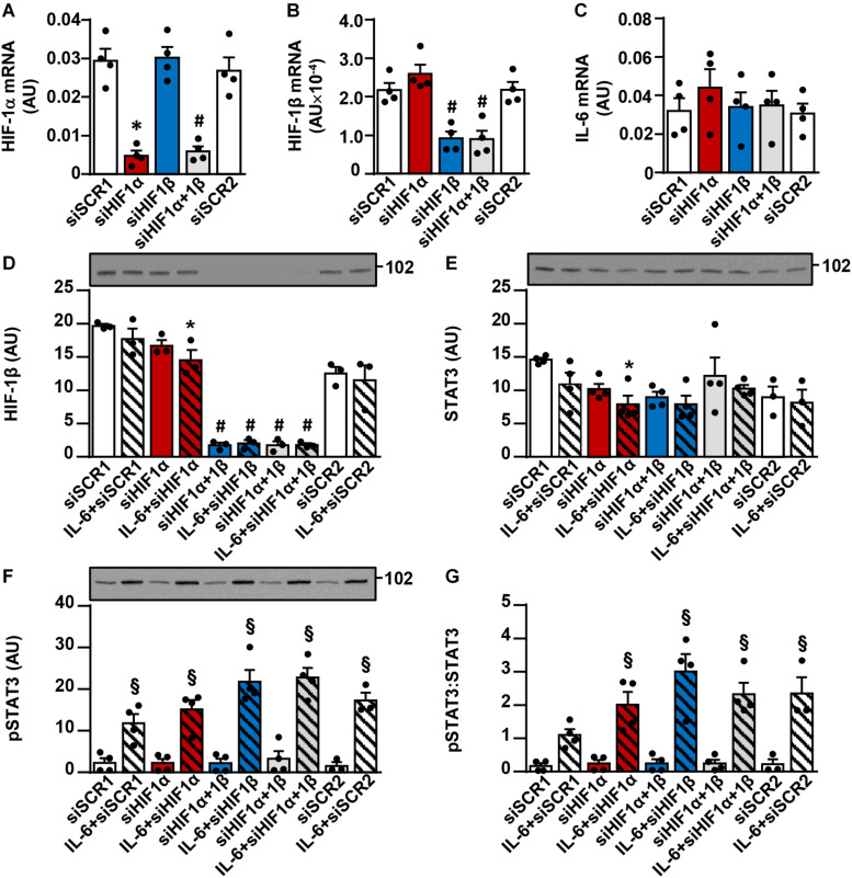
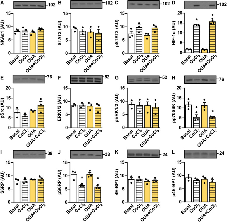
The cardiotonic steroids (CTS), such as ouabain and marinobufagenin, are thought to be adrenocortical hormones secreted during exercise and the stress response. The catalytic α-subunit of Na,K-ATPase (NKA) is a CTS receptor, whose largest pool is located in skeletal muscles, indicating that muscles are a major target for CTS. Skeletal muscles contribute to adaptations to exercise by secreting interleukin-6 (IL-6) and plethora of other cytokines, which exert paracrine and endocrine effects in muscles and non-muscle tissues. Here, we determined whether ouabain, a prototypical CTS, modulates IL-6 signaling and secretion in the cultured human skeletal muscle cells. Ouabain (2.5-50 nM) suppressed the abundance of STAT3, a key transcription factor downstream of the IL-6 receptor, as well as its basal and IL-6-stimulated phosphorylation. Conversely, ouabain (50 nM) increased the phosphorylation of ERK1/2, Akt, p70S6K, and S6 ribosomal protein, indicating activation of the ERK1/2 and the Akt-mTOR pathways. Proteasome inhibitor MG-132 blocked the ouabain-induced suppression of the total STAT3, but did not prevent the dephosphorylation of STAT3. Ouabain (50 nM) suppressed hypoxia-inducible factor-1α (HIF-1α), a modulator of STAT3 signaling, but gene silencing of HIF-1α and/or its partner protein HIF-1β did not mimic effects of ouabain on the phosphorylation of STAT3. Ouabain (50 nM) failed to suppress the phosphorylation of STAT3 and HIF-1α in rat L6 skeletal muscle cells, which express the ouabain-resistant α1-subunit of NKA. We also found that ouabain (100 nM) promoted the secretion of IL-6, IL-8, GM-CSF, and TNF-α from the skeletal muscle cells of healthy subjects, and the secretion of GM-CSF from cells of subjects with the type 2 diabetes. Marinobufagenin (10 nM), another important CTS, did not alter the secretion of these cytokines. In conclusion, our study shows that ouabain suppresses the IL-6 signaling via STAT3, but promotes the secretion of IL-6 and other cytokines, which might represent a negative feedback in the IL-6/STAT3 pathway. Collectively, our results implicate a role for CTS and NKA in regulation of the IL-6 signaling and secretion in skeletal muscle.
强心甾体(CTS),如哇巴因和海蟾蜍毒配基,被认为是运动和应激反应过程中分泌的肾上腺皮质激素。钠钾ATP酶(NKA)的催化α亚基是一种CTS受体,其最大的储存库位于骨骼肌中,这表明肌肉是CTS的主要作用靶点。骨骼肌通过分泌白细胞介素-6(IL-6)和大量其他细胞因子来促进对运动的适应,这些细胞因子在肌肉和非肌肉组织中发挥旁分泌和内分泌作用。在此,我们确定了典型的CTS——哇巴因是否能调节培养的人骨骼肌细胞中IL-6的信号传导和分泌。哇巴因(2.5 - 50 nM)抑制了IL-6受体下游关键转录因子STAT3的丰度,以及其基础磷酸化和IL-6刺激后的磷酸化。相反,哇巴因(50 nM)增加了ERK1/2、Akt、p70S6K和S6核糖体蛋白的磷酸化,表明ERK1/2和Akt - mTOR通路被激活。蛋白酶体抑制剂MG - 132阻断了哇巴因诱导的总STAT3的抑制,但并未阻止STAT3的去磷酸化。哇巴因(50 nM)抑制了缺氧诱导因子-1α(HIF - 1α),后者是STAT3信号传导的调节剂,但HIF - 1α及其伴侣蛋白HIF - 1β的基因沉默并未模拟哇巴因对STAT3磷酸化的影响。哇巴因(50 nM)未能抑制表达对哇巴因耐药的NKAα1亚基的大鼠L6骨骼肌细胞中STAT3和HIF - 1α的磷酸化。我们还发现,哇巴因(100 nM)促进了健康受试者骨骼肌细胞中IL-6、IL-8、粒细胞-巨噬细胞集落刺激因子(GM - CSF)和肿瘤坏死因子-α(TNF - α)的分泌,以及2型糖尿病患者细胞中GM - CSF的分泌。另一种重要的CTS——海蟾蜍毒配基(10 nM)并未改变这些细胞因子的分泌。总之,我们的研究表明,哇巴因通过STAT3抑制IL-6信号传导,但促进IL-6和其他细胞因子的分泌,这可能代表了IL-6/STAT3通路中的一种负反馈。总体而言,我们的结果表明CTS和NKA在调节骨骼肌中IL-6信号传导和分泌方面发挥作用。