Laboratory of Molecular Biophysics, Biochemistry and Biophysics Center, NHLBI, National Institutes of Health, Bethesda, Maryland, USA.
Electron Microscopy Core Facility, NHLBI, National Institutes of Health, Bethesda, Maryland, USA.
J Biol Chem. 2020 Dec 25;295(52):18226-18238. doi: 10.1074/jbc.RA120.013023. Epub 2020 Oct 26.
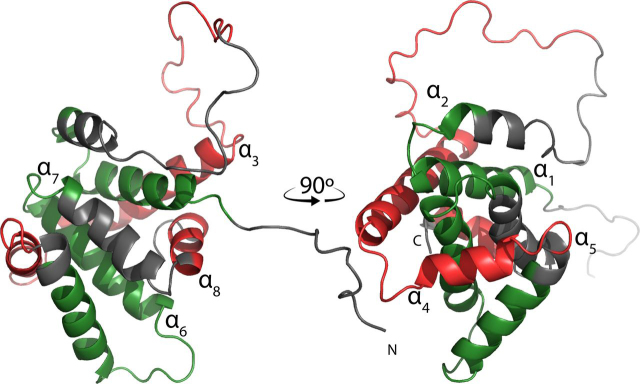
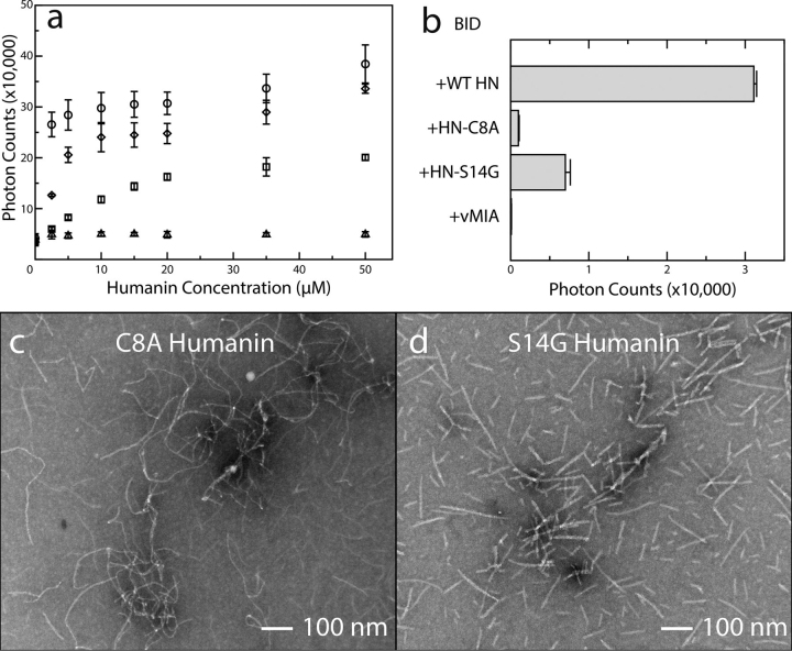
Members of the B-cell lymphoma (BCL-2) protein family regulate mitochondrial outer membrane permeabilization (MOMP), a phenomenon in which mitochondria become porous and release death-propagating complexes during the early stages of apoptosis. Pro-apoptotic BCL-2 proteins oligomerize at the mitochondrial outer membrane during MOMP, inducing pore formation. Of current interest are endogenous factors that can inhibit pro-apoptotic BCL-2 mitochondrial outer membrane translocation and oligomerization. A mitochondrial-derived peptide, Humanin (HN), was reported being expressed from an alternate ORF in the mitochondrial genome and inhibiting apoptosis through interactions with the pro-apoptotic BCL-2 proteins. Specifically, it is known to complex with BAX and BID. We recently reported the fibrillation of HN and BAX into β-sheets. Here, we detail the fibrillation between HN and BID. These fibers were characterized using several spectroscopic techniques, protease fragmentation with mass analysis, and EM. Enhanced fibrillation rates were detected with rising temperatures or pH values and the presence of a detergent. BID fibers are similar to those produced using BAX; however, the structures differ in final conformations of the BCL-2 proteins. BID fibers display both types of secondary structure in the fiber, whereas BAX was converted entirely to β-sheets. The data show that two distinct segments of BID are incorporated into the fiber structure, whereas other portions of BID remain solvent-exposed and retain helical structure. Similar analyses show that anti-apoptotic BCL-x does not form fibers with humanin. These results support a general mechanism of sequestration of pro-apoptotic BCL-2 proteins into fibers by HN to inhibit MOMP.
BCL-2 蛋白家族成员调节线粒体外膜通透性(MOMP),即在凋亡早期,线粒体变得多孔并释放促进死亡的复合物。促凋亡 BCL-2 蛋白在 MOMP 期间在线粒体外膜上寡聚,诱导孔形成。目前感兴趣的是可以抑制促凋亡 BCL-2 线粒体外膜易位和寡聚的内源性因素。一种线粒体衍生肽,人源神经保护因子(HN),据报道是从线粒体基因组中的另一个 ORF 表达的,并通过与促凋亡 BCL-2 蛋白相互作用来抑制细胞凋亡。具体来说,它与 BAX 和 BID 复合物。我们最近报道了 HN 和 BAX 纤维化为 β-片层。在这里,我们详细描述了 HN 和 BID 之间的纤维形成。使用几种光谱技术、蛋白酶片段化和质量分析以及 EM 对这些纤维进行了表征。随着温度或 pH 值的升高以及去污剂的存在,检测到增强的纤维形成速率。BID 纤维类似于使用 BAX 产生的纤维;然而,在 BCL-2 蛋白的最终构象上,这些结构有所不同。BID 纤维在纤维中显示出两种类型的二级结构,而 BAX 则完全转化为 β-片层。数据表明,BID 的两个不同片段被纳入纤维结构,而 BID 的其他部分仍然暴露在溶剂中并保持螺旋结构。类似的分析表明,抗凋亡 BCL-x 不会与人源神经保护因子形成纤维。这些结果支持了 HN 将促凋亡 BCL-2 蛋白隔离到纤维中以抑制 MOMP 的一般机制。