• 文献检索
  • 文档翻译
  • 深度研究
  • 学术资讯
  • Suppr Zotero 插件Zotero 插件
  • 邀请有礼
  • 套餐&价格
  • 历史记录
应用&插件
Suppr Zotero 插件Zotero 插件浏览器插件Mac 客户端Windows 客户端微信小程序
定价
高级版会员购买积分包购买API积分包
服务
文献检索文档翻译深度研究API 文档MCP 服务
关于我们
关于 Suppr公司介绍联系我们用户协议隐私条款
关注我们

Suppr 超能文献

核心技术专利:CN118964589B侵权必究
粤ICP备2023148730 号-1Suppr @ 2026

文献检索

告别复杂PubMed语法,用中文像聊天一样搜索,搜遍4000万医学文献。AI智能推荐,让科研检索更轻松。

立即免费搜索

文件翻译

保留排版,准确专业,支持PDF/Word/PPT等文件格式,支持 12+语言互译。

免费翻译文档

深度研究

AI帮你快速写综述,25分钟生成高质量综述,智能提取关键信息,辅助科研写作。

立即免费体验

技术说明:一种利用磁共振成像(MRI)和计算机断层扫描(CT)提高阻止本领估计准确性的方法。

Technical Note: A methodology for improved accuracy in stopping power estimation using MRI and CT.

作者信息

Scholey Jessica E, Chandramohan Dharshan, Naren Tarun, Liu William, Larson Peder Eric Zufall, Sudhyadhom Atchar

机构信息

Department of Radiation Oncology, The University of California, San Francisco, CA, USA.

Department of Radiology and Biomedical Imaging, The University of California, San Francisco, CA, USA.

出版信息

Med Phys. 2021 Jan;48(1):342-353. doi: 10.1002/mp.14555. Epub 2020 Nov 20.

DOI:10.1002/mp.14555
PMID:33107997
原文链接:https://pmc.ncbi.nlm.nih.gov/articles/PMC8717393/
Abstract

PURPOSE

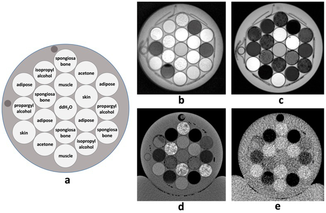
Proton therapy is becoming an increasingly popular cancer treatment modality due to the proton's physical advantage in that it deposits the majority of its energy at the distal end of its track where the tumor is located. The proton range in a material is determined from the stopping power ratio (SPR) of the material. However, SPR is typically estimated based on a computed tomography (CT) scan which can lead to range estimation errors due to the difference in x-ray and proton interactions in matter, which can preclude the ability to utilize protons to their full potential. Applications of magnetic resonance imaging (MRI) in radiotherapy have increased over the past decade and using MRI to calculate SPR directly could provide numerous advantages. The purpose of this study was to develop a practical implementation of a novel multimodal imaging method for estimating SPR and compare the results of this method to physical measurements in which values were computed directly using tissue substitute materials fabricated to mimic skin, muscle, adipose, and spongiosa bone.

METHODS

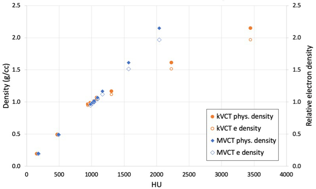
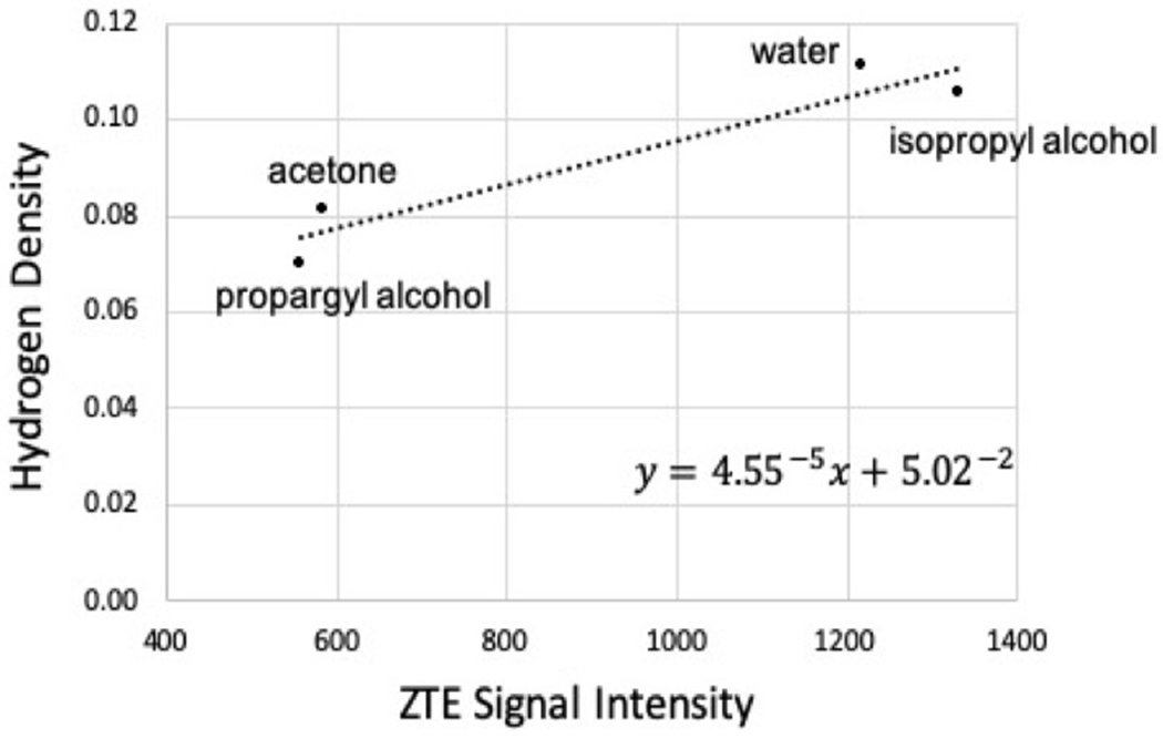
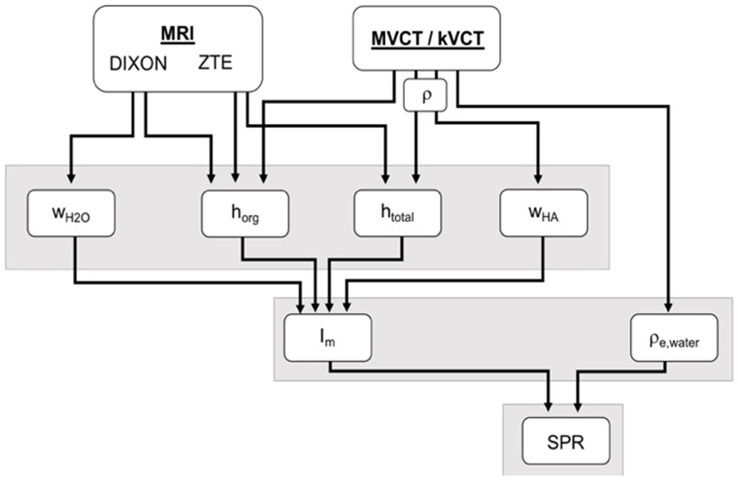
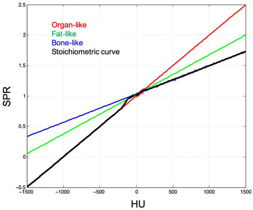
For both the multimodal imaging method and physical measurements, SPR was calculated using the Bethe-Bloch equation from values of relative electron density and mean ionization potential determined for each tissue. Parameters used to estimate SPR using the multimodal imaging method were extracted from Dixon water-only and ( H) proton density-weighted zero echo time MRI sequences and CT, with both kVCT and MVCT used separately to evaluate the performance of each. For comparison, SPR was also computed from kVCT using the stoichiometric method, the current clinical standard.

RESULTS

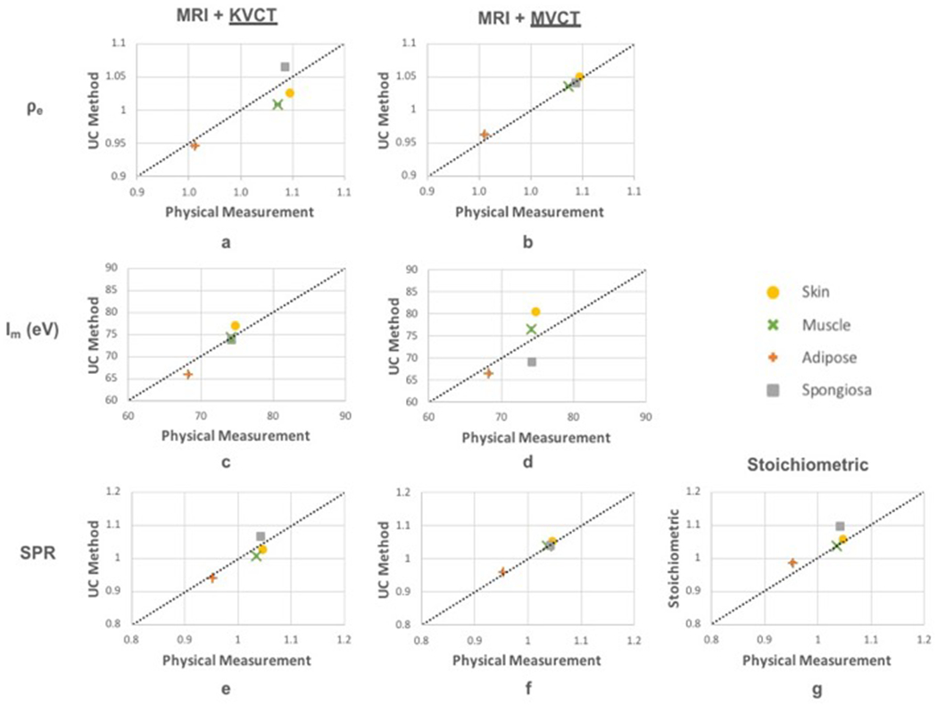
Results showed that our multimodal imaging approach using MRI with either kVCT or MVCT was in close agreement to SPR calculated from physical measurements for the four tissue substitutes evaluated. Using MRI and MVCT, SPR values estimated using our method were within 1% of physical measurements and were more accurate than the stoichiometric method for the tissue types studied.

CONCLUSIONS

We have demonstrated the methodology for improved estimation of SPR using the proposed multimodal imaging framework.

摘要

目的

质子治疗正成为一种越来越受欢迎的癌症治疗方式,这是因为质子具有物理优势,即它将大部分能量沉积在其径迹的远端,也就是肿瘤所在的位置。材料中的质子射程由该材料的阻止本领比(SPR)确定。然而,SPR通常是基于计算机断层扫描(CT)来估计的,由于X射线与质子在物质中的相互作用存在差异,这可能会导致射程估计误差,进而可能妨碍充分发挥质子的治疗潜力。在过去十年中,磁共振成像(MRI)在放射治疗中的应用有所增加,直接使用MRI来计算SPR可能会带来诸多优势。本研究的目的是开发一种用于估计SPR的新型多模态成像方法的实际实施方案,并将该方法的结果与通过使用模拟皮肤、肌肉、脂肪和松质骨制造的组织替代材料直接计算得到的物理测量值进行比较。

方法

对于多模态成像方法和物理测量,均使用贝特 - 布洛赫方程根据为每个组织确定的相对电子密度和平均电离势的值来计算SPR。使用多模态成像方法估计SPR所使用的参数是从仅水的狄克逊序列和(氢)质子密度加权零回波时间MRI序列以及CT中提取的,分别使用千伏CT(kVCT)和兆伏CT(MVCT)来评估各自的性能。为作比较,SPR也使用化学计量法从kVCT计算得出,这是当前的临床标准方法。

结果

结果表明,我们使用kVCT或MVCT的MRI多模态成像方法与针对所评估的四种组织替代物通过物理测量计算得到的SPR非常吻合。使用MRI和MVCT时,我们的方法估计的SPR值与物理测量值相差在1%以内,并且对于所研究的组织类型比化学计量法更准确。

结论

我们已经证明了使用所提出的多模态成像框架改进SPR估计的方法。

https://cdn.ncbi.nlm.nih.gov/pmc/blobs/ee12/8717393/8dcb24ce81a7/nihms-1765681-f0006.jpg
https://cdn.ncbi.nlm.nih.gov/pmc/blobs/ee12/8717393/69d701fbb05f/nihms-1765681-f0001.jpg
https://cdn.ncbi.nlm.nih.gov/pmc/blobs/ee12/8717393/b0bbb6769907/nihms-1765681-f0002.jpg
https://cdn.ncbi.nlm.nih.gov/pmc/blobs/ee12/8717393/b97bea0942dd/nihms-1765681-f0003.jpg
https://cdn.ncbi.nlm.nih.gov/pmc/blobs/ee12/8717393/6d193e2d5788/nihms-1765681-f0004.jpg
https://cdn.ncbi.nlm.nih.gov/pmc/blobs/ee12/8717393/f3b17a1a5a95/nihms-1765681-f0005.jpg
https://cdn.ncbi.nlm.nih.gov/pmc/blobs/ee12/8717393/8dcb24ce81a7/nihms-1765681-f0006.jpg

相似文献

1
Technical Note: A methodology for improved accuracy in stopping power estimation using MRI and CT.技术说明:一种利用磁共振成像(MRI)和计算机断层扫描(CT)提高阻止本领估计准确性的方法。
Med Phys. 2021 Jan;48(1):342-353. doi: 10.1002/mp.14555. Epub 2020 Nov 20.
2
Validation of an MR-based multimodal method for molecular composition and proton stopping power ratio determination using animal tissues and tissue-mimicking phantoms.基于磁共振的多模态方法用于动物组织和组织模拟体的分子组成和质子阻止本领比测定的验证。
Phys Med Biol. 2023 Aug 28;68(17). doi: 10.1088/1361-6560/ace876.
3
Improved accuracy of relative electron density and proton stopping power ratio through CycleGAN machine learning.通过CycleGAN 机器学习提高相对电子密度和质子阻止本领比的准确性。
Phys Med Biol. 2022 May 2;67(10). doi: 10.1088/1361-6560/ac6725.
4
Feasibility of using megavoltage computed tomography to reduce proton range uncertainty: A simulation study.使用兆伏级计算机断层扫描技术降低质子射程不确定性的可行性:一项模拟研究。
J Appl Clin Med Phys. 2021 Mar;22(3):131-140. doi: 10.1002/acm2.13191. Epub 2021 Feb 20.
5
MRI-only based material mass density and relative stopping power estimation via deep learning for proton therapy: a preliminary study.基于 MRI 的质子治疗物质质量密度和相对阻止本领的深度学习估算:初步研究。
Sci Rep. 2024 May 15;14(1):11166. doi: 10.1038/s41598-024-61869-8.
6
Dosimetric comparison of stopping power calibration with dual-energy CT and single-energy CT in proton therapy treatment planning.质子治疗计划中双能CT与单能CT在阻止本领校准方面的剂量学比较。
Med Phys. 2016 Jun;43(6):2845-2854. doi: 10.1118/1.4948683.
7
Stopping-power ratio estimation for proton radiotherapy using dual-energy computed tomography and prior-image constrained denoising.使用双能计算机断层扫描和先验图像约束去噪技术进行质子放疗的阻止本领比估计。
Med Phys. 2023 Mar;50(3):1481-1495. doi: 10.1002/mp.16063. Epub 2022 Nov 17.
8
Electron density and effective atomic number estimation in a maximum a posteriori framework for dual-energy computed tomography.基于最大后验概率框架的双能计算机断层扫描中的电子密度和有效原子序数估计
Med Phys. 2020 Sep;47(9):4137-4149. doi: 10.1002/mp.14309. Epub 2020 Jul 13.
9
Prediction of proton stopping power ratios using dual-energy CT basis material decomposition.使用双能CT基物质分解预测质子阻止本领比
Med Phys. 2024 Feb;51(2):881-897. doi: 10.1002/mp.16929. Epub 2024 Jan 9.
10
Synthetic dual-energy CT for MRI-only based proton therapy treatment planning using label-GAN.基于标签生成对抗网络的 MRI 单模态质子治疗计划的合成双能 CT。
Phys Med Biol. 2021 Mar 9;66(6):065014. doi: 10.1088/1361-6560/abe736.

引用本文的文献

1
Validation of dual-energy CT-based composition analysis using fresh animal tissues and composition-optimized tissue equivalent samples.采用新鲜动物组织和成分优化的组织等效样本对基于双能 CT 的成分分析进行验证。
Phys Med Biol. 2024 Aug 12;69(16):165033. doi: 10.1088/1361-6560/ad68bc.
2
MRI-only based material mass density and relative stopping power estimation via deep learning for proton therapy: a preliminary study.基于 MRI 的质子治疗物质质量密度和相对阻止本领的深度学习估算:初步研究。
Sci Rep. 2024 May 15;14(1):11166. doi: 10.1038/s41598-024-61869-8.
3
Validation of an MR-based multimodal method for molecular composition and proton stopping power ratio determination using animal tissues and tissue-mimicking phantoms.

本文引用的文献

1
Quantitative imaging for radiotherapy purposes.放射治疗的定量成像。
Radiother Oncol. 2020 May;146:66-75. doi: 10.1016/j.radonc.2020.01.026. Epub 2020 Feb 27.
2
Systematic analysis of the impact of imaging noise on dual-energy CT-based proton stopping power ratio estimation.基于双能 CT 的质子阻止本领比估计中成像噪声影响的系统分析。
Med Phys. 2019 May;46(5):2251-2263. doi: 10.1002/mp.13493. Epub 2019 Apr 1.
3
Revisiting the single-energy CT calibration for proton therapy treatment planning: a critical look at the stoichiometric method.
基于磁共振的多模态方法用于动物组织和组织模拟体的分子组成和质子阻止本领比测定的验证。
Phys Med Biol. 2023 Aug 28;68(17). doi: 10.1088/1361-6560/ace876.
4
Validation of prompt gamma-ray spectroscopy for proton range verification in tissue-mimicking and porcine samples.用于组织模拟和猪样本中质子射程验证的瞬发伽马射线光谱学的验证。
Phys Med Biol. 2022 Oct 12;67(20). doi: 10.1088/1361-6560/ac950f.
5
Generation of synthetic megavoltage CT for MRI-only radiotherapy treatment planning using a 3D deep convolutional neural network.使用三维深度卷积神经网络生成仅用于 MRI 的放疗治疗计划的合成兆伏 CT。
Med Phys. 2022 Oct;49(10):6622-6634. doi: 10.1002/mp.15876. Epub 2022 Aug 8.
6
Improved accuracy of relative electron density and proton stopping power ratio through CycleGAN machine learning.通过CycleGAN 机器学习提高相对电子密度和质子阻止本领比的准确性。
Phys Med Biol. 2022 May 2;67(10). doi: 10.1088/1361-6560/ac6725.
7
On the molecular relationship between Hounsfield Unit (HU), mass density, and electron density in computed tomography (CT).关于计算机断层扫描(CT)中亨氏单位(HU)、质量密度和电子密度之间的分子关系。
PLoS One. 2020 Dec 31;15(12):e0244861. doi: 10.1371/journal.pone.0244861. eCollection 2020.
重新审视质子治疗计划的单能量 CT 校准:化学计量法的批判性探讨。
Phys Med Biol. 2018 Nov 26;63(23):235011. doi: 10.1088/1361-6560/aaede5.
4
The Use of Quantitative Imaging in Radiation Oncology: A Quantitative Imaging Network (QIN) Perspective.定量影像学在放射肿瘤学中的应用:定量影像学网络(QIN)视角。
Int J Radiat Oncol Biol Phys. 2018 Nov 15;102(4):1219-1235. doi: 10.1016/j.ijrobp.2018.06.023. Epub 2018 Jun 30.
5
RF coils: A practical guide for nonphysicists.射频线圈:非物理学家实用指南。
J Magn Reson Imaging. 2018 Jun 13;48(3):590-604. doi: 10.1002/jmri.26187.
6
Determination of mean ionization potential using magnetic resonance imaging for the reduction of proton beam range uncertainties: theory and application.利用磁共振成像测定平均电离势以减少质子束射程不确定性:理论与应用
Phys Med Biol. 2017 Oct 27;62(22):8521-8535. doi: 10.1088/1361-6560/aa8d9e.
7
Comprehensive analysis of proton range uncertainties related to stopping-power-ratio estimation using dual-energy CT imaging.使用双能CT成像对与阻止本领比估计相关的质子射程不确定性进行综合分析。
Phys Med Biol. 2017 Aug 9;62(17):7056-7074. doi: 10.1088/1361-6560/aa7dc9.
8
The potential of dual-energy CT to reduce proton beam range uncertainties.双能 CT 降低质子束射程不确定性的潜力。
Med Phys. 2017 Jun;44(6):2332-2344. doi: 10.1002/mp.12215. Epub 2017 Apr 21.
9
A New Strategy for Fast MRI-Based Quantification of the Myelin Water Fraction: Application to Brain Imaging in Infants.一种基于快速 MRI 的髓鞘水分数定量的新策略:在婴儿脑成像中的应用。
PLoS One. 2016 Oct 13;11(10):e0163143. doi: 10.1371/journal.pone.0163143. eCollection 2016.
10
Erratum to: Solid-State Quantitative H and P MRI of Cortical Bone in Humans.《人类皮质骨的固态定量氢和磷磁共振成像》勘误
Curr Osteoporos Rep. 2016 Aug;14(4):159-161. doi: 10.1007/s11914-016-0310-7.