Department of Ophthalmology, Casey Eye Institute, Oregon Health & Science University, Portland, Oregon, United States of America.
Department of Pharmaceutical Sciences, College of Pharmacy, Oregon State University, Portland, Oregon, United States of America.
PLoS One. 2020 Oct 29;15(10):e0241006. doi: 10.1371/journal.pone.0241006. eCollection 2020.
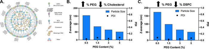
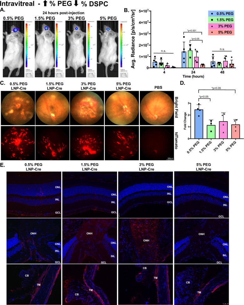
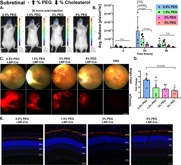
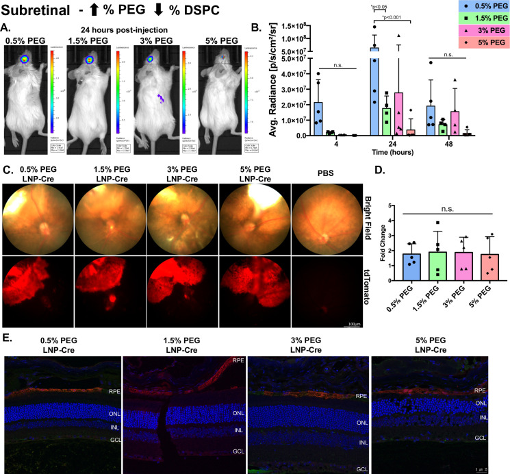
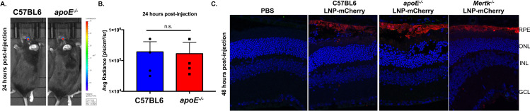
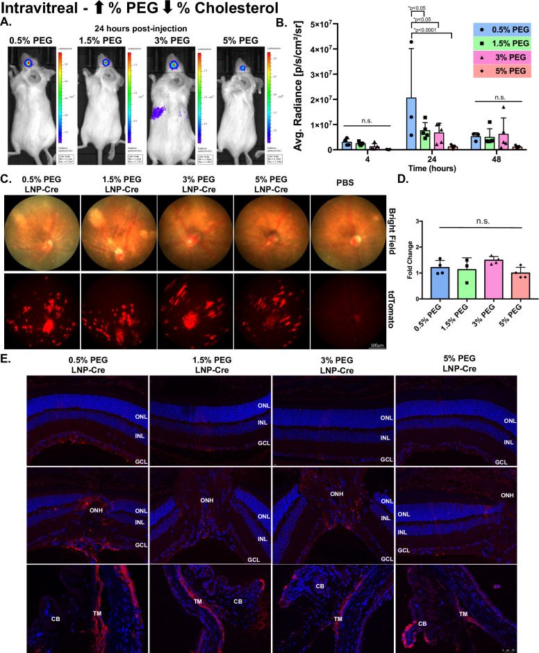
Gene therapy is now an effective approach to treat many forms of retinal degeneration. Delivery agents that are cell-specific, allow for multiple dosing regimens, and have low immunogenicity are needed to expand the utility of gene therapy for the retina. We generated eight novel lipid nanoparticles (LNPs) ranging in size from 50 nm to 150 nm by changing the PEG content from 5% to 0.5%, respectively. Subretinal injections of LNP-mRNA encoding luciferase revealed that 0.5% PEG content within nanoparticles elicits the highest expression. Similar injections of LNP delivered cre mRNA into Ai9 mice revealed cell-specific protein expression in the retinal pigment epithelium (RPE), confirmed by fundus photography and immunohistochemistry of whole globe cross-sections. To investigate mechanisms of LNP delivery to the eye, we injected mCherry mRNA using the subretinal approach in apoE-/- and Mertk-/- mice. RPE transfection was observed in both mouse models suggesting that LNP intracellular delivery is not solely dependent on apolipoprotein adsorption or phagocytosis. To investigate LNP penetration, particles were delivered to the vitreous chamber via an intravitreal injection. The 0.5% PEG particles mediated the highest luciferase activity and expression was observed in the Müller glia, the optic nerve head and the trabecular meshwork, but failed to reach the RPE. Overall, particles containing less PEG (~150 nm in size) mediated the highest expression in the eye. Thus far, these particles successfully transfect RPE, Müller cells, the optic nerve head and the trabecular meshwork based on route of administration which can expand the utility of LNP-mediated gene therapies for the eye.
基因治疗现在是治疗多种形式视网膜变性的有效方法。需要能够靶向特定细胞、允许多次给药方案且免疫原性低的递送载体,以扩大基因治疗在视网膜中的应用。我们通过将 PEG 含量分别从 5%改变到 0.5%,生成了八种大小在 50nm 到 150nm 之间的新型脂质纳米颗粒(LNPs)。通过视网膜下注射编码荧光素的 LNPs-mRNA,我们发现纳米颗粒中 0.5%PEG 含量可引发最高表达。类似地,将 LNP 递送至 Ai9 小鼠的 cre mRNA 揭示了视网膜色素上皮(RPE)中的细胞特异性蛋白表达,通过眼底照相和全眼球切片的免疫组织化学得到证实。为了研究 LNP 递送至眼睛的机制,我们通过视网膜下注射的方式在 apoE-/-和 Mertk-/-小鼠中注射了 mCherry mRNA。在这两种小鼠模型中均观察到 RPE 转染,表明 LNP 细胞内递送至眼睛不仅仅依赖于载脂蛋白吸附或吞噬作用。为了研究 LNP 的穿透性,我们通过玻璃体内注射将颗粒递送至玻璃体腔。0.5%PEG 颗粒介导了最高的荧光素酶活性和表达,在 Müller 胶质细胞、视神经头和小梁网中观察到,但是未能到达 RPE。总的来说,含有较少 PEG(~150nm 大小)的颗粒在眼睛中介导了最高的表达。到目前为止,这些颗粒根据给药途径成功转染 RPE、Müller 细胞、视神经头和小梁网,可以扩大 LNP 介导的基因治疗在眼睛中的应用。