Omarjee Loukman, Mention Pierre-Jean, Janin Anne, Kauffenstein Gilles, Pabic Estelle Le, Meilhac Olivier, Blanchard Simon, Navasiolava Nastassia, Leftheriotis Georges, Couturier Olivier, Jeannin Pascale, Lacoeuille Franck, Martin Ludovic
Vascular Medicine Department, French National Health and Medical Research (Inserm), Clinical Investigation Center (CIC) 1414, University of Rennes 1, 35033 Rennes, France.
Pseudoxanthoma Elasticum (PXE) Clinical and Research Vascular Center, CHU Rennes, 35033 Rennes, France.
J Clin Med. 2020 Oct 27;9(11):3448. doi: 10.3390/jcm9113448.
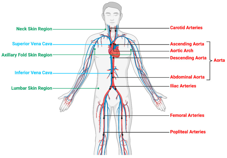
Pseudoxanthoma elasticum (PXE) is an inherited metabolic disease characterized by elastic fiber fragmentation and ectopic calcification. There is growing evidence that vascular calcification is associated with inflammatory status and is enhanced by inflammatory cytokines. Since PXE has never been considered as an inflammatory condition, no incidence of chronic inflammation leading to calcification in PXE has been reported and should be investigated. In atherosclerosis and aortic stenosis, positron emission tomography combined with computed tomographic (PET-CT) imaging has demonstrated a correlation between inflammation and calcification. The purpose of this study was to assess skin/artery inflammation and calcification in PXE patients Methods: 18F-FluroDeoxyGlucose (18F-FDG) and 18F-Sodium Fluoride (18F-NaF) PET-CT, CT-imaging and Pulse wave velocity (PWV) were used to determine skin/vascular inflammation, tissue calcification, arterial calcium score (CS) and stiffness, respectively. In addition, inorganic pyrophosphate, high-sensitive C-reactive protein and cytokines plasma levels were monitored.
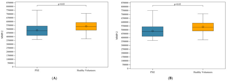
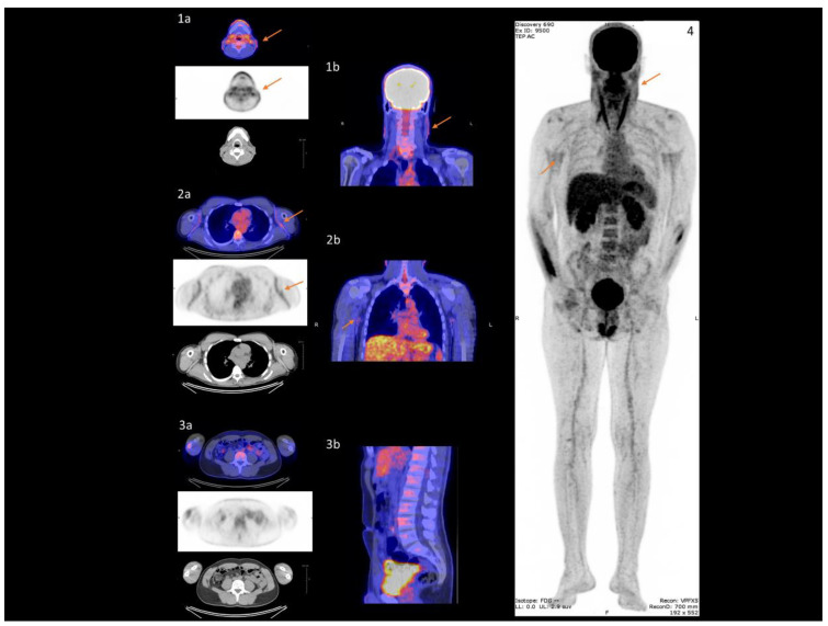
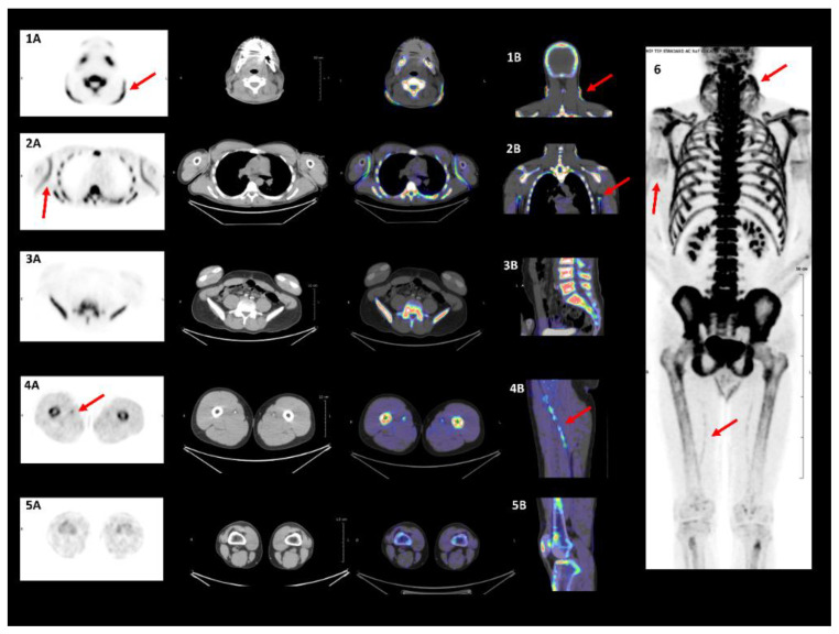
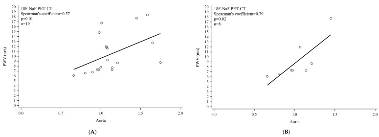
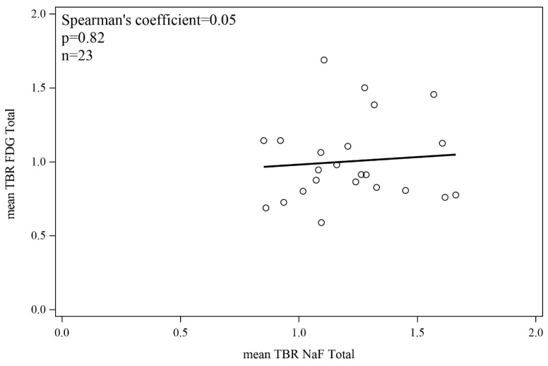
In 23 PXE patients, assessment of inflammation revealed significant 18F-FDG uptake in diseased skin areas contrary to normal regions, and exclusively in the proximal aorta contrary to the popliteal arteries. There was no correlation between 18F-FDG uptake and PWV in the aortic wall. Assessment of calcification demonstrated significant 18F-NaF uptake in diseased skin regions and in the proximal aorta and femoral arteries. 18F-NaF wall uptake correlated with CS in the femoral arteries, and aortic wall PWV. Multivariate analysis indicated that aortic wall 18F-NaF uptake is associated with diastolic blood pressure. There was no significant correlation between 18F-FDG and 18F-NaF uptake in any of the artery walls.
In the present cross-sectional study, inflammation and calcification were not correlated. PXE would appear to more closely resemble a chronic disease model of ectopic calcification than an inflammatory condition. To assess early ectopic calcification in PXE patients, 18F-NaF-PET-CT may be more relevant than CT imaging. It potentially constitutes a biomarker for disease-modifying anti-calcifying drug assessment in PXE.
弹性假黄瘤(PXE)是一种遗传性代谢疾病,其特征为弹性纤维断裂和异位钙化。越来越多的证据表明,血管钙化与炎症状态相关,且会因炎症细胞因子而加剧。由于PXE从未被视为一种炎症性疾病,因此尚未有关于PXE中导致钙化的慢性炎症发生率的报道,对此应进行研究。在动脉粥样硬化和主动脉狭窄中,正电子发射断层扫描结合计算机断层扫描(PET-CT)成像已显示出炎症与钙化之间的相关性。本研究的目的是评估PXE患者的皮肤/动脉炎症和钙化情况。方法:使用18F-氟脱氧葡萄糖(18F-FDG)和18F-氟化钠(18F-NaF)PET-CT、CT成像和脉搏波速度(PWV)分别测定皮肤/血管炎症、组织钙化、动脉钙评分(CS)和僵硬度。此外,还监测了无机焦磷酸盐、高敏C反应蛋白和细胞因子的血浆水平。
在23例PXE患者中,炎症评估显示,患病皮肤区域与正常区域相比,18F-FDG摄取显著增加,且仅在近端主动脉与腘动脉相比摄取增加。主动脉壁中18F-FDG摄取与PWV之间无相关性。钙化评估显示,患病皮肤区域、近端主动脉和股动脉中18F-NaF摄取显著增加。股动脉中18F-NaF壁摄取与CS以及主动脉壁PWV相关。多变量分析表明,主动脉壁18F-NaF摄取与舒张压相关。在任何动脉壁中,18F-FDG与18F-NaF摄取之间均无显著相关性。
在本横断面研究中,炎症与钙化不相关。PXE似乎更类似于异位钙化的慢性疾病模型,而非炎症性疾病。为了评估PXE患者的早期异位钙化,18F-NaF-PET-CT可能比CT成像更具相关性。它可能构成PXE中用于评估疾病修饰性抗钙化药物的生物标志物。