Centre for the Developing Brain, School of Imaging Sciences & Biomedical Engineering, King's College London, London, UK.
Department of Forensic and Neurodevelopmental Science, Institute of Psychiatry, Psychology and Neuroscience, King's College London, London, UK.
Neuroradiology. 2021 Apr;63(4):573-583. doi: 10.1007/s00234-020-02584-9. Epub 2020 Oct 29.
Diffusion magnetic resonance imaging (dMRI) studies report altered white matter (WM) development in preterm infants. Neurite orientation dispersion and density imaging (NODDI) metrics provide more realistic estimations of neurite architecture in vivo compared with standard diffusion tensor imaging (DTI) metrics. This study investigated microstructural maturation of WM in preterm neonates scanned between 25 and 45 weeks postmenstrual age (PMA) with normal neurodevelopmental outcomes at 2 years using DTI and NODDI metrics.
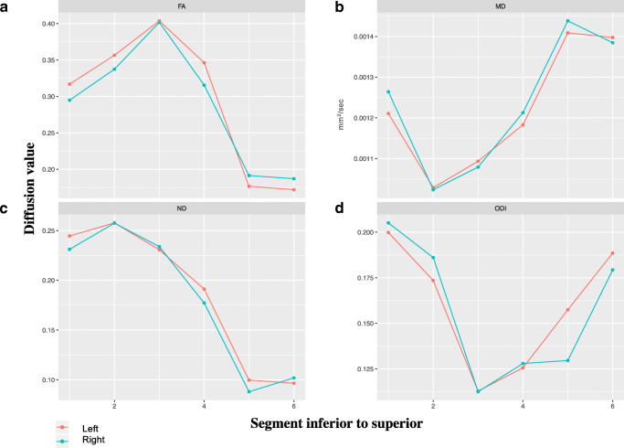
Thirty-one neonates (n = 17 male) with median (range) gestational age (GA) 32 weeks (24-36) underwent 3 T brain MRI at median (range) post menstrual age (PMA) 35 weeks (25-43). WM tracts (cingulum, fornix, corticospinal tract (CST), inferior longitudinal fasciculus (ILF), optic radiations) were delineated using constrained spherical deconvolution and probabilistic tractography in MRtrix3. DTI and NODDI metrics were extracted for the whole tract and cross-sections along each tract to assess regional development.
PMA at scan positively correlated with fractional anisotropy (FA) in the CST, fornix and optic radiations and neurite density index (NDI) in the cingulum, CST and fornix and negatively correlated with mean diffusivity (MD) in all tracts. A multilinear regression model demonstrated PMA at scan influenced all diffusion measures, GA and GAxPMA at scan influenced FA, MD and NDI and gender affected NDI. Cross-sectional analyses revealed asynchronous WM maturation within and between WM tracts.).
We describe normal WM maturation in preterm neonates with normal neurodevelopmental outcomes. NODDI can enhance our understanding of WM maturation compared with standard DTI metrics alone.
弥散磁共振成像(dMRI)研究报告早产儿的白质(WM)发育改变。与标准弥散张量成像(DTI)指标相比,神经丝取向分散和密度成像(NODDI)指标可以更真实地估计体内神经丝结构。本研究使用 DTI 和 NODDI 指标,对 25 至 45 周龄(PMA)早产儿进行扫描,在 2 岁时具有正常神经发育结局,研究 WM 的微观结构成熟。
31 例新生儿(n=17 例男性)的中位(范围)胎龄(GA)为 32 周(24-36),在中位(范围)月经龄(PMA)35 周(25-43)进行 3T 脑 MRI。使用 MRtrix3 进行约束球解卷积和概率跟踪,对 WM 束(胼胝体、穹窿、皮质脊髓束(CST)、下纵束(ILF)、视辐射)进行描绘。提取整个束和沿每个束的横截面的 DTI 和 NODDI 指标,以评估区域发育。
扫描时的 PMA 与 CST、穹窿和视辐射的各向异性分数(FA)以及胼胝体、CST 和穹窿的神经丝密度指数(NDI)呈正相关,与所有束的平均弥散度(MD)呈负相关。多元线性回归模型表明,扫描时的 PMA 影响所有扩散指标,GA 和扫描时的 GAxPMA 影响 FA、MD 和 NDI,性别影响 NDI。横截面分析显示,WM 束内和束间的 WM 成熟具有异步性。
我们描述了具有正常神经发育结局的早产儿的正常 WM 成熟。与单独使用标准 DTI 指标相比,NODDI 可以增强我们对 WM 成熟的理解。