• 文献检索
  • 文档翻译
  • 深度研究
  • 学术资讯
  • Suppr Zotero 插件Zotero 插件
  • 邀请有礼
  • 套餐&价格
  • 历史记录
应用&插件
Suppr Zotero 插件Zotero 插件浏览器插件Mac 客户端Windows 客户端微信小程序
定价
高级版会员购买积分包购买API积分包
服务
文献检索文档翻译深度研究API 文档MCP 服务
关于我们
关于 Suppr公司介绍联系我们用户协议隐私条款
关注我们

Suppr 超能文献

核心技术专利:CN118964589B侵权必究
粤ICP备2023148730 号-1Suppr @ 2026

文献检索

告别复杂PubMed语法,用中文像聊天一样搜索,搜遍4000万医学文献。AI智能推荐,让科研检索更轻松。

立即免费搜索

文件翻译

保留排版,准确专业,支持PDF/Word/PPT等文件格式,支持 12+语言互译。

免费翻译文档

深度研究

AI帮你快速写综述,25分钟生成高质量综述,智能提取关键信息,辅助科研写作。

立即免费体验

OCT 血管造影术评估急性原发性闭角型青光眼患者的黄斑血管密度和中心无血管区参数。

Macular vessel density and foveal avascular zone parameters in patients after acute primary angle closure determined by OCT angiography.

机构信息

Eye Center of Xiangya Hospital, Central South University, No. 87, Xiangya Road, Kaifu District, Changsha, 410008, Hunan, China.

Hunan Key Laboratory of Ophthalmology, Changsha, 410008, Hunan, China.

出版信息

Sci Rep. 2020 Oct 30;10(1):18717. doi: 10.1038/s41598-020-73223-9.

DOI:10.1038/s41598-020-73223-9
PMID:33127916
原文链接:https://pmc.ncbi.nlm.nih.gov/articles/PMC7603312/
Abstract

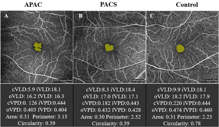
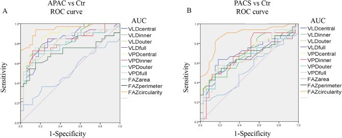
This study analyzed the optical coherence tomography angiography (OCTA) macular parameters in primary angle-closure glaucoma (PACG) patients after acute primary angle closure (APAC) episodes. Thirty-three patients with 33 APAC eyes and 33 primary angle closure suspect (PACS) eyes and 33 age-matched normal subjects (controls) were enrolled. Macular vessel density (VD) in central, inner, outer and full regions and foveal avascular zone (FAZ) parameters (area, perimeter and circularity index) were compared between APAC, PACS, and control eyes. For resolved APAC eyes, the VD in each macular region was significantly lower than that in control eyes, with less central and inner macular VD than PACS eyes. The central macular VD was significantly lower in PACS eyes than in controls. There was no difference in FAZ area and perimeter between APAC, PACS, and control eyes. FAZ circularity was highest in control eyes, followed by PACS eyes, and lowest in APAC eyes. The AUC, sensitivity and specificity of FAZ circularity were 0.944, 93.9% and 84.8%, respectively, in APAC eyes and 0.881, 84.8% and 81.8%, respectively, in PACS eyes. Therefore, FAZ circularity had the best discrimination capability for detecting both APAC and PACS eyes. Macular assessment with OCTA could provide an accurate early-stage diagnostic tool for PACG.

摘要

本研究分析了急性原发性闭角型青光眼(APAC)发作后原发性闭角型青光眼(PACG)患者的光学相干断层扫描血管造影(OCTA)黄斑参数。纳入 33 例 APAC 眼 33 例原发性闭角型青光眼可疑(PACS)眼和 33 例年龄匹配的正常对照者。比较 APAC、PACS 和对照组的黄斑中心、内、外和全区域血管密度(VD)和中心凹无血管区(FAZ)参数(面积、周长和环形度指数)。对于缓解的 APAC 眼,各黄斑区的 VD 明显低于对照组,且中心和内黄斑 VD 明显低于 PACS 眼。PACS 眼的中心黄斑 VD 明显低于对照组。APAC、PACS 和对照组的 FAZ 面积和周长无差异。FAZ 环形度在对照组最高,PACS 眼次之,APAC 眼最低。FAZ 环形度在 APAC 眼的 AUC、敏感性和特异性分别为 0.944、93.9%和 84.8%,在 PACS 眼的 AUC、敏感性和特异性分别为 0.881、84.8%和 81.8%。因此,FAZ 环形度对检测 APAC 和 PACS 眼具有最佳的判别能力。OCTA 对黄斑的评估可为 PACG 提供一种准确的早期诊断工具。

https://cdn.ncbi.nlm.nih.gov/pmc/blobs/c592/7603312/4ffc84910745/41598_2020_73223_Fig3_HTML.jpg
https://cdn.ncbi.nlm.nih.gov/pmc/blobs/c592/7603312/fc4806d936cb/41598_2020_73223_Fig1_HTML.jpg
https://cdn.ncbi.nlm.nih.gov/pmc/blobs/c592/7603312/22add03afead/41598_2020_73223_Fig2_HTML.jpg
https://cdn.ncbi.nlm.nih.gov/pmc/blobs/c592/7603312/4ffc84910745/41598_2020_73223_Fig3_HTML.jpg
https://cdn.ncbi.nlm.nih.gov/pmc/blobs/c592/7603312/fc4806d936cb/41598_2020_73223_Fig1_HTML.jpg
https://cdn.ncbi.nlm.nih.gov/pmc/blobs/c592/7603312/22add03afead/41598_2020_73223_Fig2_HTML.jpg
https://cdn.ncbi.nlm.nih.gov/pmc/blobs/c592/7603312/4ffc84910745/41598_2020_73223_Fig3_HTML.jpg

相似文献

1
Macular vessel density and foveal avascular zone parameters in patients after acute primary angle closure determined by OCT angiography.OCT 血管造影术评估急性原发性闭角型青光眼患者的黄斑血管密度和中心无血管区参数。
Sci Rep. 2020 Oct 30;10(1):18717. doi: 10.1038/s41598-020-73223-9.
2
Quantitative optical coherence tomography angiography of macular vascular structure and foveal avascular zone in glaucoma.青光眼黄斑血管结构及中心凹无血管区的定量光学相干断层扫描血管造影
PLoS One. 2017 Sep 21;12(9):e0184948. doi: 10.1371/journal.pone.0184948. eCollection 2017.
3
Changes of Optic Disc and Macular Vessel Perfusion Density in Primary Angle Closure Glaucoma: A Quantitative Study Using Optical Coherence Tomography Angiograph.原发性闭角型青光眼的视盘和黄斑血管灌注密度变化:应用光学相干断层扫描血管造影的定量研究。
Ophthalmic Res. 2023;66(1):1245-1253. doi: 10.1159/000533874. Epub 2023 Aug 30.
4
[Correlation of capillary plexus with visual acuity in idiopathic macular epiretinal membrane eyes using optical coherence tomography angiography].[利用光学相干断层扫描血管造影术研究特发性黄斑视网膜前膜眼中毛细血管丛与视力的相关性]
Zhonghua Yan Ke Za Zhi. 2019 Oct 11;55(10):757-762. doi: 10.3760/cma.j.issn.0412-4081.2019.10.006.
5
Changes of macular blood flow and structure in acute primary angle closure glaucoma.急性原发性闭角型青光眼的黄斑血流和结构变化。
Int Ophthalmol. 2022 Dec;42(12):3789-3801. doi: 10.1007/s10792-022-02399-y. Epub 2022 Jul 4.
6
Macular vessels density in diabetic retinopathy: quantitative assessment using optical coherence tomography angiography.糖尿病视网膜病变中的黄斑血管密度:使用光学相干断层扫描血管造影术进行定量评估。
Int Ophthalmol. 2019 Aug;39(8):1845-1859. doi: 10.1007/s10792-018-1013-0. Epub 2018 Sep 7.
7
Retina Vascular Structures near the Optic Disc and in the Macula in Primary Angle-Closure Suspects.原发性闭角型青光眼可疑患者视盘及黄斑区附近的视网膜血管结构
Ophthalmic Res. 2022;65(5):575-583. doi: 10.1159/000520030. Epub 2021 Oct 14.
8
Foveal avascular zone and macular vessel density after correction for magnification error in unilateral amblyopia using optical coherence tomography angiography.应用光学相干断层扫描血管造影术矫正单侧弱视的放大误差后黄斑中心凹无血管区和黄斑血管密度。
BMC Ophthalmol. 2019 Aug 5;19(1):171. doi: 10.1186/s12886-019-1177-z.
9
Quantification of Retinal Microvascular Density Using Optic Coherence Tomography Angiography in Primary Angle Closure Disease.应用光相干断层扫描血管成像定量分析原发性闭角型青光眼的视网膜血管密度。
Curr Eye Res. 2021 Jul;46(7):1018-1024. doi: 10.1080/02713683.2020.1849728. Epub 2020 Nov 26.
10
Reproducibility of Foveal Avascular Zone and Superficial Macular Retinal Vasculature Measurements in Healthy Eyes Determined by Two Different Scanning Protocols of Optical Coherence Tomography Angiography.两种不同的光学相干断层扫描血管造影扫描方案在健康眼中对中心凹无血管区和浅层视网膜脉络膜血管测量的重复性。
Ophthalmic Res. 2020;63(3):244-251. doi: 10.1159/000503071. Epub 2019 Oct 16.

引用本文的文献

1
Foveal Avascular Zone Enlargement as a Risk Factor for Early Retinal Ganglion Cell Dysfunction in Glaucoma Suspects.黄斑无血管区扩大作为青光眼可疑患者早期视网膜神经节细胞功能障碍的危险因素。
Diagnostics (Basel). 2025 Aug 21;15(16):2103. doi: 10.3390/diagnostics15162103.
2
Macular vascular density changes in different stages of chronic primary angle-closure glaucoma.慢性原发性闭角型青光眼不同阶段的黄斑血管密度变化
Front Med (Lausanne). 2025 Jul 24;12:1620673. doi: 10.3389/fmed.2025.1620673. eCollection 2025.
3
Ocular perfusion in primary angle-closure suspect and primary angle-closure eyes without prior acute angle closure.

本文引用的文献

1
Prevalence and causes of vision loss in East Asia in 2015: magnitude, temporal trends and projections.2015 年东亚视力丧失的患病率和病因:规模、时间趋势和预测。
Br J Ophthalmol. 2020 May;104(5):616-622. doi: 10.1136/bjophthalmol-2018-313308. Epub 2019 Aug 28.
2
Systemic Determinants of Peripapillary Vessel Density in Healthy African Americans: The African American Eye Disease Study.健康非裔美国人视乳头旁血管密度的系统性决定因素:非裔美国人眼病研究。
Am J Ophthalmol. 2019 Nov;207:240-247. doi: 10.1016/j.ajo.2019.06.014. Epub 2019 Jun 21.
3
Changes in vessel density of the patients with narrow antenior chamber after an acute intraocular pressure elevation observed by OCT angiography.
原发性闭角型青光眼可疑患者及既往无急性闭角型青光眼发作的原发性闭角型青光眼患者的眼灌注情况
Oman J Ophthalmol. 2025 Jun 24;18(2):133-137. doi: 10.4103/ojo.ojo_154_24. eCollection 2025 May-Aug.
4
Structural measurements and vessel density of spectral-domain optic coherence tomography in early, moderate, and severe primary angle-closure glaucoma.早期、中度和重度原发性闭角型青光眼的光谱域光学相干断层扫描的结构测量与血管密度
Int J Ophthalmol. 2023 Jul 18;16(7):1100-1109. doi: 10.18240/ijo.2023.07.15. eCollection 2023.
5
Alterations in Macular Microvasculature in Pterygium Patients Measured by OCT Angiography.通过光学相干断层扫描血管造影术测量翼状胬肉患者黄斑部微血管的改变。
Diagnostics (Basel). 2023 Apr 30;13(9):1603. doi: 10.3390/diagnostics13091603.
6
EFEMP1 is a potential biomarker of choroid thickness change in myopia.EFEMP1是近视中脉络膜厚度变化的一个潜在生物标志物。
Front Neurosci. 2023 Feb 20;17:1144421. doi: 10.3389/fnins.2023.1144421. eCollection 2023.
7
Body shape and risk of glaucoma: A Mendelian randomization.体型与青光眼风险:一项孟德尔随机化研究
Front Med (Lausanne). 2022 Sep 23;9:999974. doi: 10.3389/fmed.2022.999974. eCollection 2022.
8
Quantitative Measurements of Vessel Density and Blood Flow Areas Primary Angle Closure Diseases: A Study of Optical Coherence Tomography Angiography.原发性闭角型疾病中血管密度和血流面积的定量测量:光学相干断层扫描血管造影研究
J Clin Med. 2022 Jul 13;11(14):4040. doi: 10.3390/jcm11144040.
9
Spatial positional relationship between macular superficial vessel density and ganglion cell-inner plexiform layer thickness in primary angle closure glaucoma.原发性闭角型青光眼黄斑浅层血管密度与神经节细胞-内丛状层厚度的空间位置关系
Int Ophthalmol. 2022 Jan;42(1):103-112. doi: 10.1007/s10792-021-02005-7. Epub 2021 Aug 15.
OCT 血管造影观察急性眼压升高后窄房角患者血管密度的变化。
BMC Ophthalmol. 2019 Jun 21;19(1):132. doi: 10.1186/s12886-019-1146-6.
4
Diurnal change of retinal vessel density and mean ocular perfusion pressure in patients with open-angle glaucoma.开角型青光眼患者视网膜血管密度和平均眼灌注压的昼夜变化。
PLoS One. 2019 Apr 26;14(4):e0215684. doi: 10.1371/journal.pone.0215684. eCollection 2019.
5
Outflow facility and extent of angle closure in a porcine model.猪模型中的房水流出功能及房角关闭程度
Graefes Arch Clin Exp Ophthalmol. 2019 Jun;257(6):1239-1245. doi: 10.1007/s00417-019-04279-1. Epub 2019 Apr 3.
6
Topographic correlation between macular superficial microvessel density and ganglion cell-inner plexiform layer thickness in glaucoma-suspect and early normal-tension glaucoma.青光眼可疑患者和早期正常眼压性青光眼的黄斑浅层微血管密度与神经节细胞-内丛状层厚度的拓扑相关性。
Br J Ophthalmol. 2020 Jan;104(1):104-109. doi: 10.1136/bjophthalmol-2018-313732. Epub 2019 Apr 2.
7
Macula Vessel Density and Foveal Avascular Zone Parameters in Exfoliation Glaucoma Compared to Primary Open-Angle Glaucoma.与原发性开角型青光眼相比,原发性开角型青光眼的黄斑血管密度和中心无血管区参数。
Invest Ophthalmol Vis Sci. 2019 Mar 1;60(4):1244-1253. doi: 10.1167/iovs.18-25986.
8
Quantitative analysis of retinal and choroid capillary ischaemia using optical coherence tomography angiography in type 2 diabetes.2 型糖尿病患者视网膜和脉络膜毛细血管缺血的光相干断层扫描血管造影定量分析。
Acta Ophthalmol. 2019 May;97(3):240-246. doi: 10.1111/aos.14076. Epub 2019 Feb 27.
9
Changes in Optic Nerve Head Vessel Density After Acute Primary Angle Closure Episode.急性原发性闭角型青光眼发作后视乳头血管密度的变化。
Invest Ophthalmol Vis Sci. 2019 Feb 1;60(2):552-558. doi: 10.1167/iovs.18-25915.
10
Choroidal Microvascular Dropout in Primary Angle Closure Glaucoma.原发性闭角型青光眼的脉络膜小血管缺失。
Am J Ophthalmol. 2019 Mar;199:184-192. doi: 10.1016/j.ajo.2018.11.021. Epub 2018 Dec 12.