• 文献检索
  • 文档翻译
  • 深度研究
  • 学术资讯
  • Suppr Zotero 插件Zotero 插件
  • 邀请有礼
  • 套餐&价格
  • 历史记录
应用&插件
Suppr Zotero 插件Zotero 插件浏览器插件Mac 客户端Windows 客户端微信小程序
定价
高级版会员购买积分包购买API积分包
服务
文献检索文档翻译深度研究API 文档MCP 服务
关于我们
关于 Suppr公司介绍联系我们用户协议隐私条款
关注我们

Suppr 超能文献

核心技术专利:CN118964589B侵权必究
粤ICP备2023148730 号-1Suppr @ 2026

文献检索

告别复杂PubMed语法,用中文像聊天一样搜索,搜遍4000万医学文献。AI智能推荐,让科研检索更轻松。

立即免费搜索

文件翻译

保留排版,准确专业,支持PDF/Word/PPT等文件格式,支持 12+语言互译。

免费翻译文档

深度研究

AI帮你快速写综述,25分钟生成高质量综述,智能提取关键信息,辅助科研写作。

立即免费体验

使用 CT 脑衰减变化估计缺血性脑卒中发病时间的可行性和诊断准确性。

Feasibility and diagnostic accuracy of using brain attenuation changes on CT to estimate time of ischemic stroke onset.

机构信息

Edinburgh Imaging, and UK Dementia Research Institute at the University of Edinburgh and Centre for Clinical Brain Sciences, University of Edinburgh, Chancellor's Building, 49 Little France Crescent, Edinburgh, EH16 4SB, UK.

Centre for Clinical Brain Sciences, University of Edinburgh, Edinburgh, UK.

出版信息

Neuroradiology. 2021 Jun;63(6):869-878. doi: 10.1007/s00234-020-02591-w. Epub 2020 Oct 30.

DOI:10.1007/s00234-020-02591-w
PMID:33128140
原文链接:https://pmc.ncbi.nlm.nih.gov/articles/PMC8128810/
Abstract

PURPOSE

CT attenuation of ischemic brain reduces with time after stroke onset. We aimed to quantify this relationship and test the feasibility and accuracy of estimating stroke onset time using only CT attenuation of visible ischemic lesions, the CT-Clock Tool.

METHODS

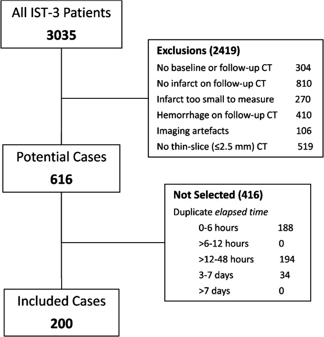
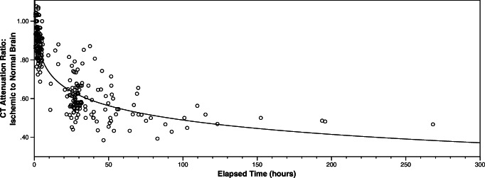
We selected CT scans with ischemic lesions representing a range of stroke-onset-to-scan times (elapsed time) from a well-defined stroke trial. We measured the attenuation of ischemic lesions and contralateral normal brain to derive attenuation ratio. We assigned scans to development (75%) or test (25%) datasets. We plotted the relationship between attenuation ratio and elapsed time in the development dataset and derived a best-fit curve. We calculated estimated time in the test dataset using only the attenuation ratio curve. We compared estimated time to elapsed time and derived absolute error for estimated time. We assessed area under the receiver operating characteristic (AUROC) curve for identifying scans ≤ 4.5 h elapsed time.

RESULTS

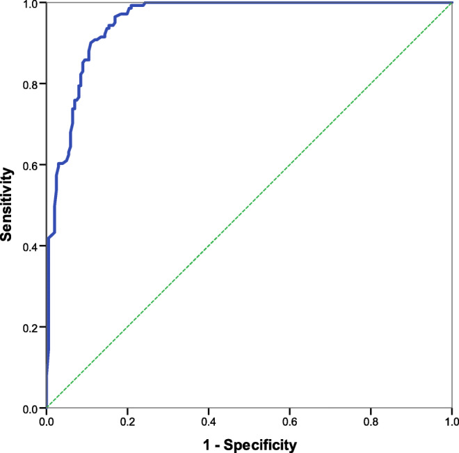
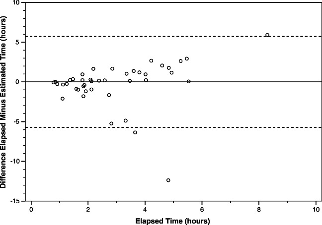
We included 342 scans from 200 patients (41% male, median age 83 years). Elapsed time range: 22 min to 36 days. Estimation errors were least at early elapsed times (r = 0.82, p < 0.0001): median absolute error was 23, 106, 1030 and 1933 min for scans acquired ≤ 3, > 3-9, > 9-30 and > 30 h from stroke onset, respectively. AUROC was high at 0.955.

CONCLUSIONS

It is feasible to accurately estimate stroke onset time using simple attenuation measures of ischemic brain. Our method was most accurate 0-9 h from onset and may be useful for treatment eligibility assessment, especially where imaging resources are limited.

摘要

目的

脑缺血的 CT 衰减值随卒中发病后时间的推移而降低。我们旨在量化这种关系,并测试仅使用可见缺血性病变的 CT 衰减值(CT-Clock 工具)来估计卒中发病时间的可行性和准确性。

方法

我们从一项明确的卒中试验中选择了具有一系列卒中发病至扫描时间(延迟时间)的 CT 扫描,这些扫描都有缺血性病变。我们测量了缺血性病变和对侧正常脑的衰减值,以得出衰减比值。我们将扫描分为开发(75%)和测试(25%)数据集。我们在开发数据集中绘制衰减比值与延迟时间之间的关系,并得出最佳拟合曲线。我们仅使用衰减比值曲线在测试数据集中计算估计时间。我们将估计时间与实际延迟时间进行比较,并计算估计时间的绝对误差。我们评估了用于识别 ≤ 4.5 小时延迟时间的扫描的接收者操作特征(ROC)曲线下面积(AUROC)。

结果

我们纳入了 200 名患者的 342 次扫描(41%为男性,中位年龄为 83 岁)。延迟时间范围:22 分钟至 36 天。在早期延迟时间,估计误差最小(r=0.82,p<0.0001):扫描获得 ≤ 3、>3-9、>9-30 和 >30 小时后,中位数绝对误差分别为 23、106、1030 和 1933 分钟。AUROC 非常高,为 0.955。

结论

使用缺血性脑的简单衰减测量值准确估计卒中发病时间是可行的。我们的方法在发病后 0-9 小时内最准确,可能对治疗资格评估有用,尤其是在影像资源有限的情况下。

https://cdn.ncbi.nlm.nih.gov/pmc/blobs/aec8/8128810/75cdcae162a5/234_2020_2591_Fig5_HTML.jpg
https://cdn.ncbi.nlm.nih.gov/pmc/blobs/aec8/8128810/9ce124fc8631/234_2020_2591_Fig1_HTML.jpg
https://cdn.ncbi.nlm.nih.gov/pmc/blobs/aec8/8128810/7958f5774936/234_2020_2591_Fig2_HTML.jpg
https://cdn.ncbi.nlm.nih.gov/pmc/blobs/aec8/8128810/09b25ac738aa/234_2020_2591_Fig3_HTML.jpg
https://cdn.ncbi.nlm.nih.gov/pmc/blobs/aec8/8128810/f0a1a6819e50/234_2020_2591_Fig4_HTML.jpg
https://cdn.ncbi.nlm.nih.gov/pmc/blobs/aec8/8128810/75cdcae162a5/234_2020_2591_Fig5_HTML.jpg

相似文献

1
Feasibility and diagnostic accuracy of using brain attenuation changes on CT to estimate time of ischemic stroke onset.使用 CT 脑衰减变化估计缺血性脑卒中发病时间的可行性和诊断准确性。
Neuroradiology. 2021 Jun;63(6):869-878. doi: 10.1007/s00234-020-02591-w. Epub 2020 Oct 30.
2
Assessing Brain Tissue Viability on Nonenhanced Computed Tomography After Ischemic Stroke.评估缺血性脑卒中后非增强 CT 上的脑组织活力。
Stroke. 2023 Feb;54(2):558-566. doi: 10.1161/STROKEAHA.122.041241. Epub 2023 Jan 5.
3
Feasibility of CT attenuation values in distinguishing acute ischemic stroke, old cerebral infarction and leukoaraiosis.CT 衰减值鉴别急性缺血性脑卒中、陈旧性脑梗死和脑白质疏松症的可行性。
BMC Med Imaging. 2024 Jun 26;24(1):160. doi: 10.1186/s12880-024-01340-2.
4
Attenuation Changes in ASPECTS Regions: A Surrogate for CT Perfusion-based Ischemic Core in Acute Ischemic Stroke.ASPECTS 区域的衰减变化:急性缺血性脑卒中 CT 灌注缺血核心的替代指标。
Radiology. 2019 May;291(2):451-458. doi: 10.1148/radiol.2019182041. Epub 2019 Mar 19.
5
A Novel Imaging Technique (X-Map) to Identify Acute Ischemic Lesions Using Noncontrast Dual-Energy Computed Tomography.一种使用非增强双能量计算机断层扫描识别急性缺血性病变的新型成像技术(X-Map)。
J Stroke Cerebrovasc Dis. 2017 Jan;26(1):34-41. doi: 10.1016/j.jstrokecerebrovasdis.2016.08.025. Epub 2016 Sep 14.
6
Performance of e-ASPECTS software in comparison to that of stroke physicians on assessing CT scans of acute ischemic stroke patients.e-ASPECTS 软件在评估急性缺血性脑卒中患者 CT 扫描方面的表现与脑卒中医师的表现比较。
Int J Stroke. 2016 Jun;11(4):438-45. doi: 10.1177/1747493016632244. Epub 2016 Feb 15.
7
Cerebral attenuation on single-phase CT angiography source images: Automated ischemia detection and morphologic outcome prediction after thrombectomy in patients with ischemic stroke.单相 CT 血管造影源图像上的脑衰减:缺血性卒中患者血栓切除术后自动缺血检测和形态学结局预测。
PLoS One. 2020 Aug 13;15(8):e0236956. doi: 10.1371/journal.pone.0236956. eCollection 2020.
8
Effect of extended CT perfusion acquisition time on ischemic core and penumbra volume estimation in patients with acute ischemic stroke due to a large vessel occlusion.延长CT灌注采集时间对大血管闭塞所致急性缺血性脑卒中患者缺血核心区和半暗带体积估计的影响。
PLoS One. 2015 Mar 19;10(3):e0119409. doi: 10.1371/journal.pone.0119409. eCollection 2015.
9
A quantitative symmetry-based analysis of hyperacute ischemic stroke lesions in noncontrast computed tomography.基于定量对称性的非增强计算机断层扫描中超急性缺血性中风病变分析
Med Phys. 2017 Jan;44(1):192-199. doi: 10.1002/mp.12015. Epub 2017 Jan 8.
10
A Quantitative Method Using Head Noncontrast CT Scans to Detect Hyperacute Nonvisible Ischemic Changes in Patients With Stroke.一种使用头部非增强CT扫描检测中风患者超急性隐匿性缺血性改变的定量方法。
J Neuroimaging. 2016 Nov;26(6):581-587. doi: 10.1111/jon.12363. Epub 2016 May 30.

引用本文的文献

1
Characteristics of peripheral immune response induced by large-vessel occlusion in patients with acute ischemic stroke.急性缺血性脑卒中患者大血管闭塞诱导的外周免疫反应特征
Front Neurol. 2024 Dec 10;15:1512720. doi: 10.3389/fneur.2024.1512720. eCollection 2024.
2
Deep learning biomarker of chronometric and biological ischemic stroke lesion age from unenhanced CT.基于平扫CT的计时性和生物性缺血性脑卒中病变年龄的深度学习生物标志物
NPJ Digit Med. 2024 Dec 6;7(1):338. doi: 10.1038/s41746-024-01325-z.
3
A dual-energy computed tomography-based radiomics nomogram for predicting time since stroke onset: a multicenter study.

本文引用的文献

1
Lesion Age Imaging in Acute Stroke: Water Uptake in CT Versus DWI-FLAIR Mismatch.急性卒中的病灶年龄影像学:CT 水摄取与 DWI-FLAIR 不匹配。
Ann Neurol. 2020 Dec;88(6):1144-1152. doi: 10.1002/ana.25903. Epub 2020 Oct 2.
2
Eligibility for late endovascular treatment using DAWN, DEFUSE-3, and more liberal selection criteria in a stroke center.在卒中中心,采用 DAWN、DEFUSE-3 及更宽松的选择标准进行血管内晚期治疗的入选标准。
J Neurointerv Surg. 2020 Sep;12(9):842-847. doi: 10.1136/neurintsurg-2019-015382. Epub 2019 Nov 26.
3
Thrombolysis Guided by Perfusion Imaging up to 9 Hours after Onset of Stroke.
基于双能 CT 的放射组学列线图预测脑卒中介入治疗后时间:一项多中心研究。
Eur Radiol. 2024 Nov;34(11):7373-7385. doi: 10.1007/s00330-024-10802-8. Epub 2024 Jun 4.
4
Diffusion-/perfusion-weighted imaging fusion to automatically identify stroke within 4.5 h.弥散/灌注加权成像融合可在 4.5 小时内自动识别中风。
Eur Radiol. 2024 Oct;34(10):6808-6819. doi: 10.1007/s00330-024-10619-5. Epub 2024 Mar 15.
5
The Scottish Medical Imaging Archive: 57.3 Million Radiology Studies Linked to Their Medical Records.苏格兰医学影像档案:5730 万份放射学研究与病历相关联。
Radiol Artif Intell. 2024 Jan;6(1):e220266. doi: 10.1148/ryai.220266.
6
Normal Appearing Ischaemic Brain Tissue on CT and Outcome After Intravenous Alteplase.CT上表现正常的缺血性脑组织及静脉注射阿替普酶后的预后
Front Radiol. 2022 Jun 22;2:902165. doi: 10.3389/fradi.2022.902165. eCollection 2022.
7
Assessing Brain Tissue Viability on Nonenhanced Computed Tomography After Ischemic Stroke.评估缺血性脑卒中后非增强 CT 上的脑组织活力。
Stroke. 2023 Feb;54(2):558-566. doi: 10.1161/STROKEAHA.122.041241. Epub 2023 Jan 5.
8
A Review of Risk Factors and Predictors for Hemorrhagic Transformation in Patients with Acute Ischemic Stroke.急性缺血性脑卒中患者出血转化的危险因素及预测因素综述
Int J Vasc Med. 2021 Dec 6;2021:4244267. doi: 10.1155/2021/4244267. eCollection 2021.
发病 9 小时内采用灌注成像指导的溶栓治疗。
N Engl J Med. 2019 May 9;380(19):1795-1803. doi: 10.1056/NEJMoa1813046.
4
MRI-Guided Thrombolysis for Stroke with Unknown Time of Onset.MRI 引导下的不明时间起病脑卒中溶栓治疗。
N Engl J Med. 2018 Aug 16;379(7):611-622. doi: 10.1056/NEJMoa1804355. Epub 2018 May 16.
5
Thrombectomy for Stroke at 6 to 16 Hours with Selection by Perfusion Imaging.6至16小时卒中的血栓切除术及灌注成像选择
N Engl J Med. 2018 Feb 22;378(8):708-718. doi: 10.1056/NEJMoa1713973. Epub 2018 Jan 24.
6
Thrombectomy 6 to 24 Hours after Stroke with a Mismatch between Deficit and Infarct.发病后 6 至 24 小时内进行取栓术治疗与缺损和梗死不匹配的脑卒中。
N Engl J Med. 2018 Jan 4;378(1):11-21. doi: 10.1056/NEJMoa1706442. Epub 2017 Nov 11.
7
Fast Versus Slow Progressors of Infarct Growth in Large Vessel Occlusion Stroke: Clinical and Research Implications.大血管闭塞性卒中梗死灶生长的快速进展者与缓慢进展者:临床及研究意义
Stroke. 2017 Sep;48(9):2621-2627. doi: 10.1161/STROKEAHA.117.017673. Epub 2017 Aug 9.
8
Imaging of cerebral ischemic edema and neuronal death.脑缺血性水肿与神经元死亡的影像学检查
Neuroradiology. 2017 Jun;59(6):545-553. doi: 10.1007/s00234-017-1847-6. Epub 2017 May 24.
9
ASPECTS (Alberta Stroke Program Early CT Score) Measurement Using Hounsfield Unit Values When Selecting Patients for Stroke Thrombectomy.在为卒中取栓术选择患者时,使用亨氏单位值进行ASPECTS(阿尔伯塔卒中项目早期CT评分)测量。
Stroke. 2017 Jun;48(6):1574-1579. doi: 10.1161/STROKEAHA.117.016745. Epub 2017 May 9.
10
Computed tomography-based quantification of lesion water uptake identifies patients within 4.5 hours of stroke onset: A multicenter observational study.基于计算机断层扫描的病灶水摄取定量分析可在卒中发病后 4.5 小时内识别患者:一项多中心观察性研究。
Ann Neurol. 2016 Dec;80(6):924-934. doi: 10.1002/ana.24818.