• 文献检索
  • 文档翻译
  • 深度研究
  • 学术资讯
  • Suppr Zotero 插件Zotero 插件
  • 邀请有礼
  • 套餐&价格
  • 历史记录
应用&插件
Suppr Zotero 插件Zotero 插件浏览器插件Mac 客户端Windows 客户端微信小程序
定价
高级版会员购买积分包购买API积分包
服务
文献检索文档翻译深度研究API 文档MCP 服务
关于我们
关于 Suppr公司介绍联系我们用户协议隐私条款
关注我们

Suppr 超能文献

核心技术专利:CN118964589B侵权必究
粤ICP备2023148730 号-1Suppr @ 2026

文献检索

告别复杂PubMed语法,用中文像聊天一样搜索,搜遍4000万医学文献。AI智能推荐,让科研检索更轻松。

立即免费搜索

文件翻译

保留排版,准确专业,支持PDF/Word/PPT等文件格式,支持 12+语言互译。

免费翻译文档

深度研究

AI帮你快速写综述,25分钟生成高质量综述,智能提取关键信息,辅助科研写作。

立即免费体验

在局灶节段性肾小球硬化和微小病变性肾病患者中肾小球脏层上皮细胞的原位评估。

In situ evaluation of podocytes in patients with focal segmental glomerulosclerosis and minimal change disease.

机构信息

Department of Pathology, Genetics and Evolution, Discipline of General Pathology, Institute of Biological and Natural Sciences, Federal University of Triângulo Mineiro, Uberaba, Minas Gerais, Brazil.

出版信息

PLoS One. 2020 Nov 4;15(11):e0241745. doi: 10.1371/journal.pone.0241745. eCollection 2020.

DOI:10.1371/journal.pone.0241745
PMID:33147279
原文链接:https://pmc.ncbi.nlm.nih.gov/articles/PMC7641434/
Abstract

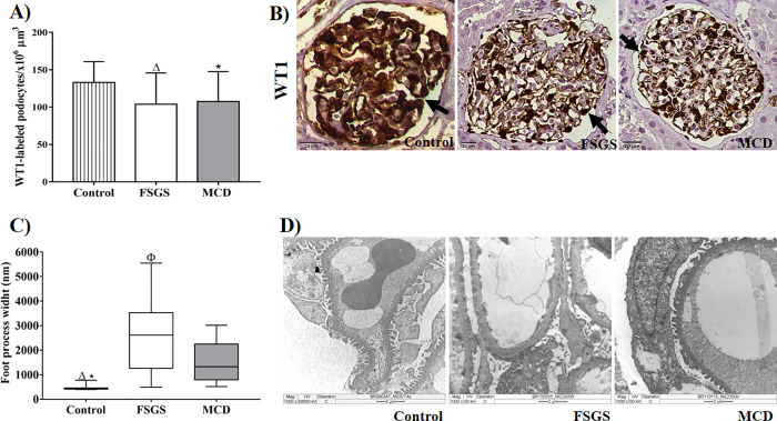
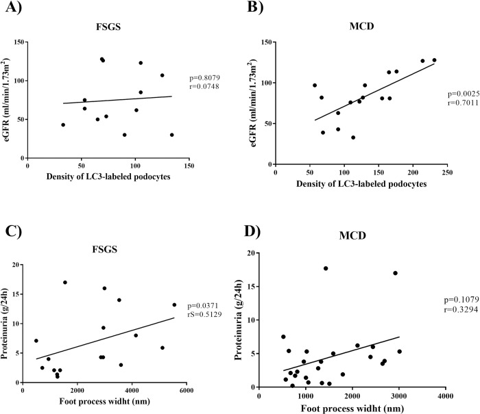
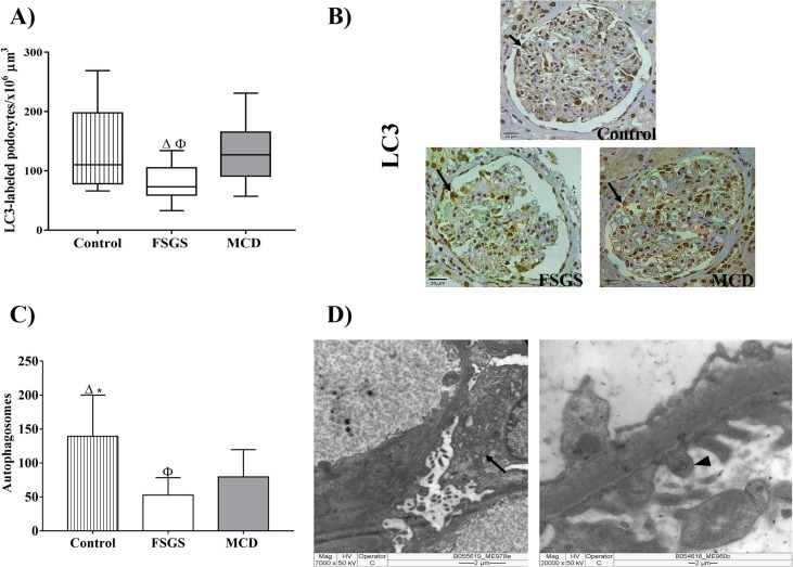
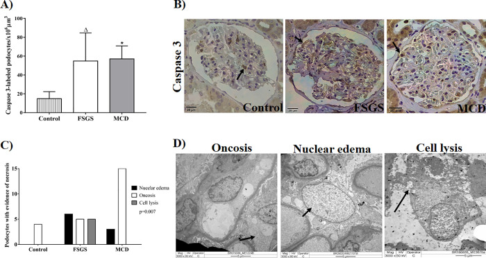
Podocyte injury in focal segmental glomerulosclerosis (FSGS) and minimal change disease (MCD) results from the imbalance between adaptive responses that maintain homeostasis and cellular dysfunction that can culminate in cell death. Therefore, an in situ analysis was performed to detect morphological changes related to cell death and autophagy in renal biopsies from adult patients with podocytopathies. Forty-nine renal biopsies from patients with FSGS (n = 22) and MCD (n = 27) were selected. In situ expression of Wilms Tumor 1 protein (WT1), light chain microtubule 1-associated protein (LC3) and caspase-3 protein were evaluated by immunohistochemistry. The foot process effacement and morphological alterations related to podocyte cell death and autophagy were analyzed with transmission electronic microscopy. Reduction in the density of WT1-labeled podocytes was observed for FSGS and MCD cases as compared to controls. Foot process width (FPW) in control group was lower than in cases of podocytopathies. In FSGS group, FPW was significantly higher than in MCD group and correlated with proteinuria. A density of LC3-labeled podocytes and the number of autophagosomes in podocytes/ pedicels were higher in the MCD group than in the FSGS group. The number of autophagosomes correlated positively with the estimated glomerular filtration rate in cases of MCD. The density of caspase-3-labeled podocytes in FSGS and MCD was higher than control group, and a higher number of podocytes with an evidence of necrosis was detected in FSGS cases than in MCD and control cases. Podocytes from patients diagnosed with FSGS showed more morphological and functional alterations resulting from a larger number of lesions and reduced cell adaptation.

摘要

足细胞损伤在局灶节段性肾小球硬化症(FSGS)和微小病变性肾病(MCD)中是由于维持内稳态的适应性反应和可能导致细胞死亡的细胞功能障碍之间的失衡所致。因此,进行了原位分析以检测与足细胞病变患者肾活检中细胞死亡和自噬相关的形态变化。选择了 49 例 FSGS(n = 22)和 MCD(n = 27)患者的肾活检。通过免疫组织化学评估 Wilms 瘤 1 蛋白(WT1)、轻链微管 1 相关蛋白(LC3)和半胱氨酸蛋白酶-3 蛋白的原位表达。用透射电子显微镜分析足突融合和与足细胞细胞死亡和自噬相关的形态改变。与对照组相比,FSGS 和 MCD 病例的 WT1 标记的足细胞密度降低。对照组的足突宽度(FPW)低于足细胞病变组。在 FSGS 组中,FPW 显著高于 MCD 组,与蛋白尿相关。MCD 组的 LC3 标记的足细胞密度和足细胞/ pedicels 中的自噬体数量高于 FSGS 组。MCD 病例中的自噬体数量与估计的肾小球滤过率呈正相关。FSGS 和 MCD 病例中 caspase-3 标记的足细胞密度高于对照组,并且在 FSGS 病例中检测到比 MCD 和对照组更多的具有坏死证据的足细胞。FSGS 诊断的患者的足细胞表现出更多的形态和功能改变,这是由于病变数量增加和细胞适应性降低所致。

https://cdn.ncbi.nlm.nih.gov/pmc/blobs/bab0/7641434/e43f7ee16c47/pone.0241745.g004.jpg
https://cdn.ncbi.nlm.nih.gov/pmc/blobs/bab0/7641434/161b1e4adfaf/pone.0241745.g001.jpg
https://cdn.ncbi.nlm.nih.gov/pmc/blobs/bab0/7641434/2ed149118fd7/pone.0241745.g002.jpg
https://cdn.ncbi.nlm.nih.gov/pmc/blobs/bab0/7641434/b3b779b6106a/pone.0241745.g003.jpg
https://cdn.ncbi.nlm.nih.gov/pmc/blobs/bab0/7641434/e43f7ee16c47/pone.0241745.g004.jpg

相似文献

1
In situ evaluation of podocytes in patients with focal segmental glomerulosclerosis and minimal change disease.在局灶节段性肾小球硬化和微小病变性肾病患者中肾小球脏层上皮细胞的原位评估。
PLoS One. 2020 Nov 4;15(11):e0241745. doi: 10.1371/journal.pone.0241745. eCollection 2020.
2
Ultrastructural features and expression of cytoskeleton proteins of podocyte from patients with minimal change disease and focal segmental glomerulosclerosis.微小病变病和局灶节段性肾小球硬化症患者足细胞的超微结构特征及细胞骨架蛋白表达
Ren Fail. 2008;30(5):477-83. doi: 10.1080/08860220802060497.
3
Cell-cycle regulatory proteins in the podocyte in collapsing glomerulopathy in children.儿童塌陷型肾小球病中足细胞的细胞周期调节蛋白
Kidney Int. 2006 Aug;70(3):529-35. doi: 10.1038/sj.ki.5001577. Epub 2006 Jun 14.
4
Podocin and uPAR are good biomarkers in cases of Focal and segmental glomerulosclerosis in pediatric renal biopsies.足细胞和尿激酶型纤溶酶原激活物受体(uPAR)是儿童肾活检局灶节段性肾小球硬化的良好生物标志物。
PLoS One. 2019 Jun 12;14(6):e0217569. doi: 10.1371/journal.pone.0217569. eCollection 2019.
5
Podocyte autophagy is associated with foot process effacement and proteinuria in patients with minimal change nephrotic syndrome.足细胞自噬与微小病变肾病综合征患者的足突消失和蛋白尿有关。
PLoS One. 2020 Jan 24;15(1):e0228337. doi: 10.1371/journal.pone.0228337. eCollection 2020.
6
Podocyte and endothelial injury in focal segmental glomerulosclerosis: an ultrastructural analysis.局灶节段性肾小球硬化中的足细胞和内皮细胞损伤:超微结构分析
Virchows Arch. 2015 Oct;467(4):449-58. doi: 10.1007/s00428-015-1821-9. Epub 2015 Aug 13.
7
Assessment of urinary angiotensinogen as a marker of podocyte injury in proteinuric nephropathies.评估尿血管紧张素原作为蛋白尿性肾病中足细胞损伤标志物的作用。
Am J Physiol Renal Physiol. 2016 Feb 15;310(4):F322-33. doi: 10.1152/ajprenal.00260.2015. Epub 2015 Dec 2.
8
Clinical significance of fibroblast-specific protein-1 expression on podocytes in patients with focal segmental glomerulosclerosis.成纤维细胞特异性蛋白-1 在局灶节段性肾小球硬化症患者的足细胞中的表达的临床意义。
Nephron Clin Pract. 2012;120(1):c1-7. doi: 10.1159/000334184. Epub 2011 Nov 23.
9
Immunoexpression of podocyte-associated proteins in acquired human glomerulopathies with nephrotic syndrome.足细胞相关蛋白在获得性肾病综合征人类肾小球病中的免疫表达
Pol J Pathol. 2006;57(1):17-21.
10
Analysis of glomerular deposition of IgM and C3 in patients with podocytopathies.分析足细胞病患者肾小球 IgM 和 C3 的沉积。
Ann Diagn Pathol. 2024 Jun;70:152292. doi: 10.1016/j.anndiagpath.2024.152292. Epub 2024 Mar 7.

引用本文的文献

1
Machine learning based identification of anoikis related gene classification patterns and immunoinfiltration characteristics in diabetic nephropathy.基于机器学习的糖尿病肾病中失巢凋亡相关基因分类模式及免疫浸润特征的识别
Sci Rep. 2025 May 1;15(1):15271. doi: 10.1038/s41598-025-99395-w.
2
Digital pathology assessment of kidney glomerular filtration barrier ultrastructure in an animal model of podocytopathy.足细胞病动物模型中肾小球滤过屏障超微结构的数字病理学评估
Biol Methods Protoc. 2025 Mar 28;10(1):bpaf024. doi: 10.1093/biomethods/bpaf024. eCollection 2025.
3
Podocyte A20/TNFAIP3 Controls Glomerulonephritis Severity via the Regulation of Inflammatory Responses and Effects on the Cytoskeleton.

本文引用的文献

1
Podocyte autophagy is associated with foot process effacement and proteinuria in patients with minimal change nephrotic syndrome.足细胞自噬与微小病变肾病综合征患者的足突消失和蛋白尿有关。
PLoS One. 2020 Jan 24;15(1):e0228337. doi: 10.1371/journal.pone.0228337. eCollection 2020.
2
Evaluation of the Diagnostic Potential of uPAR as a Biomarker in Renal Biopsies of Patients with FSGS.评估 uPAR 作为 FSGS 患者肾活检生物标志物的诊断潜力。
Dis Markers. 2019 May 2;2019:1070495. doi: 10.1155/2019/1070495. eCollection 2019.
3
Podocin and uPAR are good biomarkers in cases of Focal and segmental glomerulosclerosis in pediatric renal biopsies.
足细胞A20/TNFAIP3通过调节炎症反应和对细胞骨架的作用来控制肾小球肾炎的严重程度。
Cells. 2025 Mar 5;14(5):381. doi: 10.3390/cells14050381.
4
Influence of Podocyte Injury on the Development of Class IV Lupus Nephritis.足细胞损伤对IV型狼疮性肾炎发展的影响。
Int J Nephrol Renovasc Dis. 2024 Oct 4;17:215-225. doi: 10.2147/IJNRD.S473616. eCollection 2024.
5
Activation of the inflammasome and pyroptosis cascade in podocytes of patients with minimal change disease.微小病变病患者足细胞中炎性小体的激活及细胞焦亡级联反应。
Clin Kidney J. 2024 Jul 16;17(8):sfae216. doi: 10.1093/ckj/sfae216. eCollection 2024 Aug.
6
The IRE1α pathway in glomerular diseases: The unfolded protein response and beyond.肾小球疾病中的IRE1α信号通路:未折叠蛋白反应及其他。
Front Mol Med. 2022 Sep 26;2:971247. doi: 10.3389/fmmed.2022.971247. eCollection 2022.
7
Digital pathology assessment of kidney glomerular filtration barrier ultrastructure in an animal model of podocytopathy.在足细胞病动物模型中对肾小球滤过屏障超微结构进行数字病理学评估。
bioRxiv. 2024 Jul 15:2024.06.14.599097. doi: 10.1101/2024.06.14.599097.
8
Kidney Aging and Chronic Kidney Disease.肾脏老化与慢性肾脏病。
Int J Mol Sci. 2024 Jun 14;25(12):6585. doi: 10.3390/ijms25126585.
9
Autophagy and mitophagy: physiological implications in kidney inflammation and diseases.自噬和线粒体自噬:在肾脏炎症和疾病中的生理意义。
Am J Physiol Renal Physiol. 2023 Jul 1;325(1):F1-F21. doi: 10.1152/ajprenal.00012.2023. Epub 2023 May 11.
10
In situ assessment of Mindin as a biomarker of podocyte lesions in diabetic nephropathy.在糖尿病肾病中作为足细胞病变生物标志物的 Mindin 的原位评估。
PLoS One. 2023 May 2;18(5):e0284789. doi: 10.1371/journal.pone.0284789. eCollection 2023.
足细胞和尿激酶型纤溶酶原激活物受体(uPAR)是儿童肾活检局灶节段性肾小球硬化的良好生物标志物。
PLoS One. 2019 Jun 12;14(6):e0217569. doi: 10.1371/journal.pone.0217569. eCollection 2019.
4
Focal Segmental Glomerulosclerosis.局灶节段性肾小球硬化症
Clin J Am Soc Nephrol. 2017 Mar 7;12(3):502-517. doi: 10.2215/CJN.05960616. Epub 2017 Feb 27.
5
How many podocyte autophagosomes are there in immunoglobulin A nephropathy and idiopathic membranous nephropathy?在免疫球蛋白A肾病和特发性膜性肾病中存在多少足细胞自噬体?
Int Urol Nephrol. 2016 Dec;48(12):2109-2114. doi: 10.1007/s11255-016-1398-5. Epub 2016 Aug 31.
6
Live or Let Die: Is There any Cell Death in Podocytes?生存还是毁灭:足细胞中存在细胞死亡现象吗?
Semin Nephrol. 2016 May;36(3):208-19. doi: 10.1016/j.semnephrol.2016.03.008.
7
Podocyte injury and its consequences.足细胞损伤及其后果。
Kidney Int. 2016 Jun;89(6):1221-30. doi: 10.1016/j.kint.2016.01.012. Epub 2016 Mar 19.
8
Glomerular Aging and Focal Global Glomerulosclerosis: A Podometric Perspective.肾小球衰老与局灶节段性肾小球硬化:足细胞计量学视角
J Am Soc Nephrol. 2015 Dec;26(12):3162-78. doi: 10.1681/ASN.2014080752. Epub 2015 Jun 2.
9
Glomerular parietal epithelial cells contribute to adult podocyte regeneration in experimental focal segmental glomerulosclerosis.肾小球壁层上皮细胞在实验性局灶节段性肾小球硬化中对成年足细胞再生有贡献。
Kidney Int. 2015 Nov;88(5):999-1012. doi: 10.1038/ki.2015.152. Epub 2015 May 20.
10
The importance of podocyte adhesion for a healthy glomerulus.足细胞黏附对健康肾小球的重要性。
Front Endocrinol (Lausanne). 2014 Oct 14;5:160. doi: 10.3389/fendo.2014.00160. eCollection 2014.