Qin Caipeng, Yin Huaqi, Liu Huixin, Liu Feng, Du Yiqing, Xu Tao
Department of Urology, Peking University People's Hospital, No. 11, Xi Zhi Men Nan Street, Beijing 100044, China.
Department of Urology, Henan Provincial People's Hospital, No. 7, Wei Wu Road, Zhengzhou 450003, China.
Diagnostics (Basel). 2020 Nov 2;10(11):895. doi: 10.3390/diagnostics10110895.
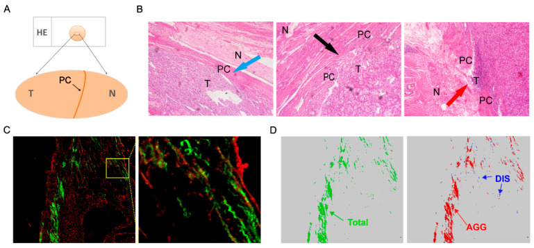
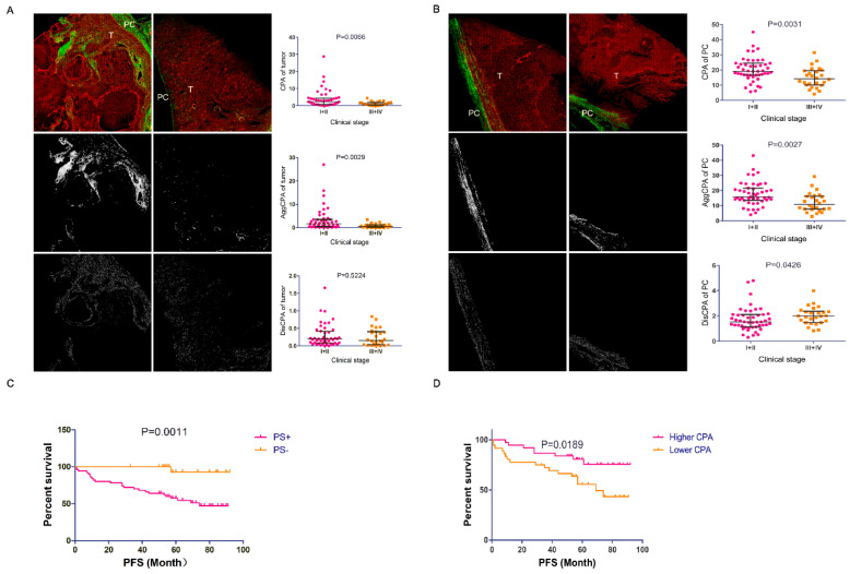
Fibrosis plays an important role in tumor growth and progression, and thus, we aimed to determine whether renal fibrosis is correlated with the clinical and pathological characteristics and prognosis of clear cell renal cell carcinoma (ccRCC). Fibrosis, including intra-tumoral fibrosis (ITF), pseudo-capsule (PC) fibrosis and adjacent normal renal interstitial fibrosis, was evaluated in 73 pairs of ccRCC specimens using second harmonic generation combined with two-photon excitation fluorescence (SHG/TPEF). The clinical and pathological characteristics of the patients who were eligible for the present study were recorded. The associations between fibrosis and clinicopathological parameters were analyzed using a Mann-Whitney U test or logistic regression analysis. Progression-free survival (PFS) was analyzed using the Kaplan-Meier method and a Cox regression model. High-resolution images of fibrosis were captured from unstained slides using the SHG/TPEF approach. Both ITF and PC fibrosis were associated with tumor progression in ccRCC. Multivariate logistic regression analysis revealed a significant inverse association between the PC collagen proportional area (CPA) and PC invasion ( < 0.05), suggesting that PC CPA is an independent risk factor or marker for PC invasion. A significant decrease in progression-free survival (PFS), determined by Kaplan-Meier curves, was observed for patients with higher PC CPA status compared with those with lower PC CPA status ( < 0.05). Similar results were observed in patients with PC invasion. In multivariate Cox regression analysis, PC invasion and intra-tumoral necrosis were identified as independent prognostic factors for PFS. Our data suggest that ITF and PC fibrosis are associated with ccRCC progression. In addition, PC fibrosis may act as a marker of PC invasion and an effective quantitative measurement for assessing prognosis.
纤维化在肿瘤生长和进展中起重要作用,因此,我们旨在确定肾纤维化是否与透明细胞肾细胞癌(ccRCC)的临床病理特征及预后相关。使用二次谐波产生结合双光子激发荧光(SHG/TPEF)技术,对73对ccRCC标本中的纤维化情况进行评估,包括肿瘤内纤维化(ITF)、假包膜(PC)纤维化和邻近正常肾间质纤维化。记录符合本研究条件的患者的临床和病理特征。采用Mann-Whitney U检验或逻辑回归分析来分析纤维化与临床病理参数之间的关联。采用Kaplan-Meier法和Cox回归模型分析无进展生存期(PFS)。使用SHG/TPEF方法从未染色的载玻片中获取纤维化的高分辨率图像。ITF和PC纤维化均与ccRCC的肿瘤进展相关。多因素逻辑回归分析显示,PC胶原比例面积(CPA)与PC侵犯之间存在显著负相关(<0.05),提示PC CPA是PC侵犯的独立危险因素或标志物。与PC CPA较低的患者相比,Kaplan-Meier曲线显示PC CPA较高的患者无进展生存期(PFS)显著降低(<0.05)。在PC侵犯的患者中也观察到类似结果。多因素Cox回归分析中,PC侵犯和肿瘤内坏死被确定为PFS的独立预后因素。我们的数据表明,ITF和PC纤维化与ccRCC进展相关。此外,PC纤维化可能作为PC侵犯的标志物以及评估预后的有效定量指标。