• 文献检索
  • 文档翻译
  • 深度研究
  • 学术资讯
  • Suppr Zotero 插件Zotero 插件
  • 邀请有礼
  • 套餐&价格
  • 历史记录
应用&插件
Suppr Zotero 插件Zotero 插件浏览器插件Mac 客户端Windows 客户端微信小程序
定价
高级版会员购买积分包购买API积分包
服务
文献检索文档翻译深度研究API 文档MCP 服务
关于我们
关于 Suppr公司介绍联系我们用户协议隐私条款
关注我们

Suppr 超能文献

核心技术专利:CN118964589B侵权必究
粤ICP备2023148730 号-1Suppr @ 2026

文献检索

告别复杂PubMed语法,用中文像聊天一样搜索,搜遍4000万医学文献。AI智能推荐,让科研检索更轻松。

立即免费搜索

文件翻译

保留排版,准确专业,支持PDF/Word/PPT等文件格式,支持 12+语言互译。

免费翻译文档

深度研究

AI帮你快速写综述,25分钟生成高质量综述,智能提取关键信息,辅助科研写作。

立即免费体验

重新评估体外氧葡萄糖剥夺脑片模型中缺氧性播散性去极化作为终末事件。

Reappraisal of anoxic spreading depolarization as a terminal event during oxygen-glucose deprivation in brain slices in vitro.

机构信息

Laboratory of Neurobiology, Kazan Federal University, Kazan, 420008, Russia.

INMED/INSERM UMR1249, Aix-Marseille University, 163 Avenue de Luminy BP13, 13273, Marseille Cedex 09, France.

出版信息

Sci Rep. 2020 Nov 4;10(1):18970. doi: 10.1038/s41598-020-75975-w.

DOI:10.1038/s41598-020-75975-w
PMID:33149164
原文链接:https://pmc.ncbi.nlm.nih.gov/articles/PMC7643106/
Abstract

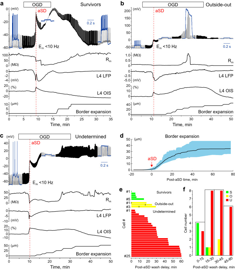
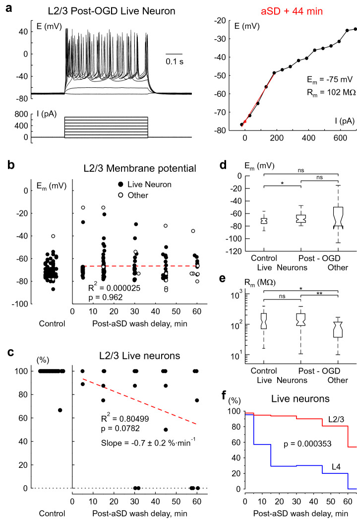
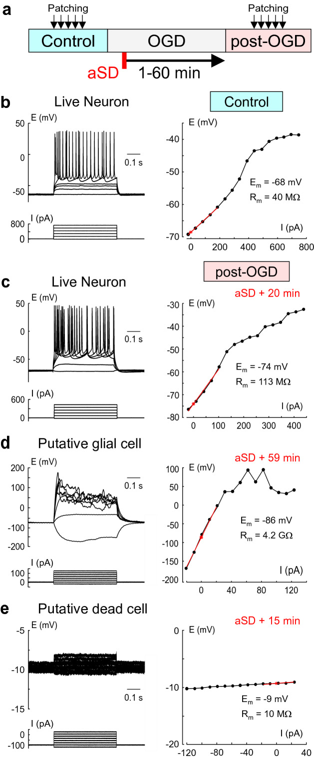
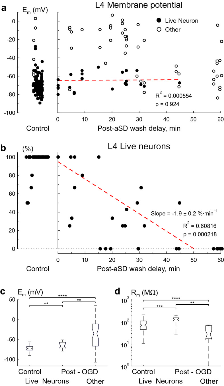
Anoxic spreading depolarization (aSD) has been hypothesized as a terminal event during oxygen-glucose deprivation (OGD) in submerged cortical slices in vitro. However, mechanical artifacts caused by aSD-triggered edema may introduce error in the assessment of neuronal viability. Here, using continuous patch-clamp recordings from submerged rat cortical slices, we first confirmed that vast majority of L4 neurons permanently lost their membrane potential during OGD-induced aSD. In some recordings, spontaneous transition from whole-cell to out-side out configuration occurred during or after aSD, and only a small fraction of neurons survived aSD with reperfusion started shortly after aSD. Secondly, to minimize artifacts caused by OGD-induced edema, cells were short-term patched following OGD episodes of various duration. Nearly half of L4 cells maintained membrane potential and showed the ability to spike-fire if reperfusion started less than 10 min after aSD. The probability of finding live neurons progressively decreased at longer reperfusion delays at a rate of about 2% per minute. We also found that neurons in L2/3 show nearly threefold higher resistance to OGD than neurons in L4. Our results suggest that in the OGD ischemia model, aSD is not a terminal event, and that the "commitment point" of irreversible damage occurs at variable delays, in the range of tens of minutes, after OGD-induced aSD in submerged cortical slices.

摘要

缺氧性弥散性去极化(aSD)被假设为体外浸没皮质切片缺氧-葡萄糖剥夺(OGD)期间的终末事件。然而,由 aSD 触发的水肿引起的机械伪影可能会在评估神经元活力时引入误差。在这里,我们使用来自浸没的大鼠皮质切片的连续膜片钳记录,首先证实,在 OGD 诱导的 aSD 期间,绝大多数 L4 神经元永久性地失去了其膜电位。在一些记录中,在 aSD 期间或之后自发地从全细胞转变为外侧向外配置,只有一小部分神经元在 aSD 后不久开始再灌注时幸存下来。其次,为了最小化 OGD 诱导的水肿引起的伪影,在 OGD 发作后对细胞进行短期贴片。如果再灌注在 aSD 后少于 10 分钟开始,近一半的 L4 细胞保持膜电位并显示出爆发的能力。如果再灌注延迟时间较长,找到存活神经元的概率以每分钟约 2%的速度逐渐降低。我们还发现 L2/3 中的神经元对 OGD 的抵抗力比 L4 中的神经元高近三倍。我们的结果表明,在 OGD 缺血模型中,aSD 不是终末事件,并且在 OGD 诱导的浸没皮质切片中的 aSD 之后,不可逆转损伤的“承诺点”以数十分钟的范围发生可变延迟。

https://cdn.ncbi.nlm.nih.gov/pmc/blobs/4e31/7643106/45643fec62c8/41598_2020_75975_Fig4_HTML.jpg
https://cdn.ncbi.nlm.nih.gov/pmc/blobs/4e31/7643106/70401684329a/41598_2020_75975_Fig1_HTML.jpg
https://cdn.ncbi.nlm.nih.gov/pmc/blobs/4e31/7643106/8d1ae62e5b55/41598_2020_75975_Fig2_HTML.jpg
https://cdn.ncbi.nlm.nih.gov/pmc/blobs/4e31/7643106/f109869af696/41598_2020_75975_Fig3_HTML.jpg
https://cdn.ncbi.nlm.nih.gov/pmc/blobs/4e31/7643106/45643fec62c8/41598_2020_75975_Fig4_HTML.jpg

相似文献

1
Reappraisal of anoxic spreading depolarization as a terminal event during oxygen-glucose deprivation in brain slices in vitro.重新评估体外氧葡萄糖剥夺脑片模型中缺氧性播散性去极化作为终末事件。
Sci Rep. 2020 Nov 4;10(1):18970. doi: 10.1038/s41598-020-75975-w.
2
Oxygen and glucose deprivation (OGD)-induced spreading depression in the Substantia Nigra.氧葡萄糖剥夺(OGD)诱导的黑质扩散性抑制。
Brain Res. 2013 Aug 21;1527:209-21. doi: 10.1016/j.brainres.2013.06.016. Epub 2013 Jun 21.
3
Anoxic spreading depolarization in the neonatal rat cortex .新生大鼠皮层中的缺氧性扩散性去极化
Front Cell Neurosci. 2023 Mar 9;17:1106268. doi: 10.3389/fncel.2023.1106268. eCollection 2023.
4
Pharmacological inhibition of the Na(+)/Ca(2+) exchanger enhances depolarizations induced by oxygen/glucose deprivation but not responses to excitatory amino acids in rat striatal neurons.对钠钙交换体的药理学抑制增强了氧糖剥夺诱导的去极化,但不影响大鼠纹状体神经元对兴奋性氨基酸的反应。
Stroke. 1999 Aug;30(8):1687-94. doi: 10.1161/01.str.30.8.1687.
5
Changes in membrane potential and the intracellular calcium concentration during CSD and OGD in layer V and layer II/III mouse cortical neurons.在 V 层和 II/III 层小鼠皮层神经元中,CSD 和 OGD 期间膜电位和细胞内钙离子浓度的变化。
J Neurophysiol. 2010 Dec;104(6):3203-12. doi: 10.1152/jn.00922.2009. Epub 2010 Sep 1.
6
Imaging anoxic depolarization during ischemia-like conditions in the mouse hemi-brain slice.在小鼠半脑切片的缺血样条件下对缺氧去极化进行成像。
J Neurophysiol. 2001 Jan;85(1):414-24. doi: 10.1152/jn.2001.85.1.414.
7
Oxygen and glucose deprivation induces major dysfunction in the somatosensory cortex of the newborn rat.氧糖剥夺会导致新生大鼠体感皮层出现严重功能障碍。
Eur J Neurosci. 2005 Nov;22(9):2295-305. doi: 10.1111/j.1460-9568.2005.04398.x.
8
Dynamics of the Hypoxia-Induced Tissue Edema in the Rat Barrel Cortex .大鼠桶状皮层缺氧诱导的组织水肿动态变化
Front Cell Neurosci. 2018 Dec 18;12:502. doi: 10.3389/fncel.2018.00502. eCollection 2018.
9
Differential spread of anoxic depolarization contributes to the pattern of neuronal injury after oxygen and glucose deprivation (OGD) in the Substantia Nigra in rat brain slices.缺氧去极化的差异传播导致大鼠脑片黑质中氧糖剥夺(OGD)后神经元损伤的模式。
Neuroscience. 2017 Jan 6;340:359-372. doi: 10.1016/j.neuroscience.2016.10.067. Epub 2016 Nov 5.
10
A novel method for inducing focal ischemia in vitro.一种新的体外诱导局灶性缺血的方法。
J Neurosci Methods. 2010 Jun 30;190(1):20-7. doi: 10.1016/j.jneumeth.2010.04.017. Epub 2010 Apr 22.

引用本文的文献

1
Enhanced Response of ZnO Nanorod-Based Flexible MEAs for Recording Ischemia-Induced Neural Activity in Acute Brain Slices.用于记录急性脑片中缺血诱导神经活动的基于氧化锌纳米棒的柔性微电极阵列的增强响应。
Nanomaterials (Basel). 2025 Jul 30;15(15):1173. doi: 10.3390/nano15151173.
2
Atypical plume-like events contribute to glutamate accumulation in metabolic stress conditions.非典型羽状样事件在代谢应激条件下导致谷氨酸积累。
iScience. 2025 Mar 20;28(4):112256. doi: 10.1016/j.isci.2025.112256. eCollection 2025 Apr 18.
3
Behavioral and Cognitive Consequences of Spreading Depolarizations: A Translational Scoping Review.

本文引用的文献

1
Cellular mechanisms underlying the rapid depolarization caused by oxygen and glucose deprivation in layer III pyramidal cells of the somatosensory cortex.体感皮层III层锥体细胞中由氧和葡萄糖剥夺引起的快速去极化的细胞机制。
Neurosci Res. 2021 Mar;164:1-9. doi: 10.1016/j.neures.2020.03.003. Epub 2020 Mar 31.
2
Restoration of brain circulation and cellular functions hours post-mortem.恢复死后数小时的大脑循环和细胞功能。
Nature. 2019 Apr;568(7752):336-343. doi: 10.1038/s41586-019-1099-1. Epub 2019 Apr 17.
3
Spreading depolarization and neuronal damage or survival in mouse neocortical brain slices immediately and 12 hours following middle cerebral artery occlusion.
扩散性去极化的行为和认知后果:一项转化性范围综述
J Neurotrauma. 2025 Jan;42(1-2):1-18. doi: 10.1089/neu.2024.0118. Epub 2024 Oct 22.
4
Brief and Diverse Excitotoxic Insults Increase the Neuronal Nuclear Membrane Permeability in the Neonatal Brain, Resulting in Neuronal Dysfunction and Cell Death.短暂而多样的兴奋性毒性损伤会增加新生脑神经元核膜通透性,导致神经元功能障碍和细胞死亡。
J Neurosci. 2024 Oct 9;44(41):e0350242024. doi: 10.1523/JNEUROSCI.0350-24.2024.
5
Trauma in Neonatal Acute Brain Slices Alters Calcium and Network Dynamics and Causes Calpain-Mediated Cell Death.新生儿急性脑片损伤改变钙和网络动力学,并导致钙蛋白酶介导的细胞死亡。
eNeuro. 2024 Jul 8;11(7). doi: 10.1523/ENEURO.0007-24.2024. Print 2024 Jul.
6
Comparative Study of Terminal Cortical Potentials Using Iridium and Ag/AgCl Electrodes.使用铱电极和 Ag/AgCl 电极的终皮质电位的对比研究。
Int J Mol Sci. 2023 Jun 28;24(13):10769. doi: 10.3390/ijms241310769.
7
Between life and death: the brain twilight zones.生死之间:大脑的边缘地带。
Front Neurosci. 2023 May 15;17:1156368. doi: 10.3389/fnins.2023.1156368. eCollection 2023.
8
Anoxic spreading depolarization in the neonatal rat cortex .新生大鼠皮层中的缺氧性扩散性去极化
Front Cell Neurosci. 2023 Mar 9;17:1106268. doi: 10.3389/fncel.2023.1106268. eCollection 2023.
9
Synaptic modifications transform neural networks to function without oxygen.突触修饰可使神经网络在无氧条件下发挥功能。
BMC Biol. 2023 Mar 16;21(1):54. doi: 10.1186/s12915-023-01518-0.
10
Depth-profile of impairments in endothelin-1 - induced focal cortical ischemia.内皮素-1 诱导局灶性皮质缺血损伤的深度分析。
J Cereb Blood Flow Metab. 2022 Oct;42(10):1944-1960. doi: 10.1177/0271678X221107422. Epub 2022 Jun 14.
大脑中动脉阻塞后即刻和 12 小时内对小鼠新皮层脑片的扩散性去极化和神经元损伤或存活的影响。
J Neurophysiol. 2019 May 1;121(5):1650-1663. doi: 10.1152/jn.00670.2018. Epub 2019 Feb 27.
4
Dynamics of the Hypoxia-Induced Tissue Edema in the Rat Barrel Cortex .大鼠桶状皮层缺氧诱导的组织水肿动态变化
Front Cell Neurosci. 2018 Dec 18;12:502. doi: 10.3389/fncel.2018.00502. eCollection 2018.
5
Preferential Initiation and Spread of Anoxic Depolarization in Layer 4 of Rat Barrel Cortex.大鼠桶状皮层第4层缺氧去极化的优先起始与传播
Front Cell Neurosci. 2017 Dec 15;11:390. doi: 10.3389/fncel.2017.00390. eCollection 2017.
6
Spreading depolarization is not an epiphenomenon but the principal mechanism of the cytotoxic edema in various gray matter structures of the brain during stroke.扩散性去极化不是一种附带现象,而是中风时大脑各种灰质结构细胞毒性水肿的主要机制。
Neuropharmacology. 2018 May 15;134(Pt B):189-207. doi: 10.1016/j.neuropharm.2017.09.027. Epub 2017 Sep 21.
7
Zika Virus Disrupts Phospho-TBK1 Localization and Mitosis in Human Neuroepithelial Stem Cells and Radial Glia.寨卡病毒扰乱人神经上皮干细胞和放射状胶质细胞中磷酸化TBK1的定位及有丝分裂。
Cell Rep. 2016 Sep 6;16(10):2576-2592. doi: 10.1016/j.celrep.2016.08.038. Epub 2016 Aug 24.
8
Large extracellular space leads to neuronal susceptibility to ischemic injury in a Na+/K+ pumps-dependent manner.大细胞外间隙以一种依赖于钠钾泵的方式导致神经元对缺血性损伤敏感。
J Comput Neurosci. 2016 Apr;40(2):177-92. doi: 10.1007/s10827-016-0591-y. Epub 2016 Feb 6.
9
Atlas of the Postnatal Rat Brain in Stereotaxic Coordinates.《立体定位坐标下的新生大鼠脑图谱》
Front Neuroanat. 2015 Dec 23;9:161. doi: 10.3389/fnana.2015.00161. eCollection 2015.
10
Molecular pathophysiology of cerebral edema.脑水肿的分子病理生理学
J Cereb Blood Flow Metab. 2016 Mar;36(3):513-38. doi: 10.1177/0271678X15617172. Epub 2015 Nov 16.