Galbusera Riccardo, Parmar Katrin, Boillat Yohan, Fartaria Mario Joao, Todea Alexandra-Ramona, Brien Kieran O', Smolinski Anna, Kappos Ludwig, van der Zwaag Wietske, Granziera Cristina
Neurologic Clinic and Policlinic, Departments of Medicine, Clinical Research and Biomedical Engineering, University Hospital Basel and University of Basel, Basel, Switzerland.
Translational Imaging in Neurology (ThINk) Basel, Department of Biomedical Engineering, University Hospital Basel and University of Basel, Basel, Switzerland.
Mult Scler J Exp Transl Clin. 2020 Oct 23;6(4):2055217320961409. doi: 10.1177/2055217320961409. eCollection 2020 Oct-Dec.
To date, little is known about the presence and extent of cerebellar cortical pathology in early stages of MS.
The aims of this study were to (i) investigate microstructural changes in the normal-appearing cerebellar cortex of early MS patients by using 7 T MRI and (ii) evaluate the influence of those changes on clinical performance.
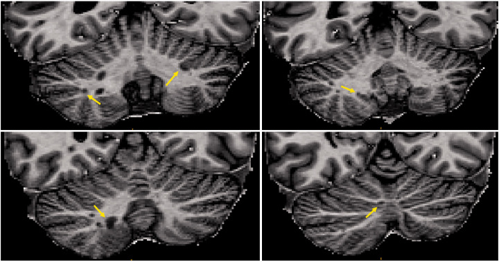
Eighteen RRMS patients and nine healthy controls underwent quantitative T and T* measurement at 7 T MRI using high-resolution MP2RAGE and multi-echo gradient-echo imaging. After subtracting lesion masks, average T and T* maps were computed for three layers in the cerebellar cortex and compared between groups using mixed effects models.
The volume of the cerebellar cortex and its layers did not differ between patients and controls. In MS patients, significantly longer T values were observed in all vermis cortical layers and in the middle and external cortical layer of the cerebellar hemispheres. No between-group differences in T* values were found. T values correlated with EDSS, SDMT and PASAT.
We found MRI evidence of damage in the normal-appearing cerebellar cortex at early MS stages and before volumetric changes. This microstructural alteration appears to be related to EDSS and cognitive performance.
迄今为止,对于多发性硬化症(MS)早期小脑皮质病变的存在情况及程度知之甚少。
本研究的目的是:(i)通过使用7T磁共振成像(MRI)研究早期MS患者外观正常的小脑皮质的微观结构变化;(ii)评估这些变化对临床表型的影响。
18例复发缓解型多发性硬化症(RRMS)患者和9名健康对照者接受了7T MRI检查,使用高分辨率MP2RAGE和多回波梯度回波成像进行定量T和T测量。在减去病变掩码后,计算小脑皮质三层的平均T和T图,并使用混合效应模型在组间进行比较。
患者和对照者之间小脑皮质及其各层的体积没有差异。在MS患者中,所有蚓部皮质层以及小脑半球的中间和外侧皮质层均观察到明显更长的T值。未发现组间T*值存在差异。T值与扩展残疾状态量表(EDSS)、符号数字模态测试(SDMT)和听觉连续加法测试(PASAT)相关。
我们发现MRI证据表明,在MS早期且在体积变化之前,外观正常的小脑皮质存在损伤。这种微观结构改变似乎与EDSS和认知表现有关。