Department of Neurology, University of Maryland School of Medicine, Baltimore, Maryland, United States of America.
University of Maryland School of Medicine, Baltimore, Maryland, United States of America.
PLoS One. 2021 Apr 26;16(4):e0249973. doi: 10.1371/journal.pone.0249973. eCollection 2021.
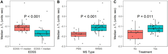
Although the blood-brain barrier (BBB) is altered in most multiple sclerosis (MS) lesions, gadolinium enhancement is seen only in acute lesions. In this study, we aimed to investigate gadolinium-induced changes in T1 relaxation time in MS lesions on 7-tesla (7T) MRI as a means to quantify BBB breakdown in non-enhancing MS lesions. Forty-seven participants with MS underwent 7T MRI of the brain with a magnitude-prepared rapid acquisition of 2 gradient echoes (MP2RAGE) sequence before and after contrast. Subtraction of pre- and post-contrast T1 maps was used to measure T1 relaxation time change (ΔT1) from gadolinium. ΔT1 values were interrogated in enhancing white matter lesions (ELs), non-enhancing white matter lesions (NELs), and normal appearing white matter (NAWM) and metrics were compared to clinical data. ΔT1 was measurable in NELs (median: -0.139 (-0.304, 0.174) seconds; p < 0.001) and was negligible in NAWM (median: -0.001 (-0.036, 0.155) seconds; p = 0.516). Median ΔT1 in NELs correlated with disability as measured by Expanded Disability Status Scale (EDSS) (rho = -0.331, p = 0.026). Multiple measures of NEL ΔT1 variability also correlated with EDSS. NEL ΔT1 values were greater and more variable in patients with progressive forms of MS and greater in those not on MS treatment. Measurement of the changes in T1 relaxation time caused by contrast on 7T MP2RAGE reveals clinically relevant evidence of BBB breakdown in NELs in MS. This data suggests that NEL ΔT1 should be evaluated further as a potential biomarker of persistently disrupted BBB in MS. Although the blood-brain barrier (BBB) is altered in most multiple sclerosis (MS) lesions, gadolinium enhancement is seen only in acute lesions. In this study, we aimed to investigate gadolinium-induced changes in T1 relaxation time in MS lesions on 7-tesla (7T) MRI as a means to quantify BBB breakdown in non-enhancing MS lesions. Forty-seven participants with MS underwent 7T MRI of the brain with a magnitude-prepared rapid acquisition of 2 gradient echoes (MP2RAGE) sequence before and after contrast. Subtraction of pre- and post-contrast T1 maps was used to measure T1 relaxation time change (ΔT1) from gadolinium. ΔT1 values were interrogated in enhancing white matter lesions (ELs), non-enhancing white matter lesions (NELs), and normal appearing white matter (NAWM) and metrics were compared to clinical data. ΔT1 was measurable in NELs (median: -0.139 (-0.304, 0.174) seconds; p < 0.001) and was negligible in NAWM (median: -0.001 (-0.036, 0.155) seconds; p = 0.516). Median ΔT1 in NELs correlated with disability as measured by Expanded Disability Status Scale (EDSS) (rho = -0.331, p = 0.026). Multiple measures of NEL ΔT1 variability also correlated with EDSS. NEL ΔT1 values were greater and more variable in patients with progressive forms of MS and greater in those not on MS treatment. Measurement of the changes in T1 relaxation time caused by contrast on 7T MP2RAGE reveals clinically relevant evidence of BBB breakdown in NELs in MS. This data suggests that NEL ΔT1 should be evaluated further as a potential biomarker of persistently disrupted BBB in MS.
虽然血脑屏障(BBB)在大多数多发性硬化症(MS)病变中发生改变,但仅在急性病变中可见钆增强。在这项研究中,我们旨在研究 7 特斯拉(7T)MRI 上 MS 病变中钆诱导的 T1 弛豫时间变化,作为量化非增强 MS 病变中 BBB 破裂的一种手段。47 名 MS 患者在对比前后接受了大脑的 7T MRI 检查,采用幅度准备快速获取 2 个梯度回波(MP2RAGE)序列。通过减去对比前后的 T1 图谱来测量来自钆的 T1 弛豫时间变化(ΔT1)。在增强的白质病变(ELs)、非增强的白质病变(NELs)和正常表现的白质(NAWM)中检查ΔT1 值,并将指标与临床数据进行比较。在 NELs 中可以测量到ΔT1(中位数:-0.139(-0.304,0.174)秒;p<0.001),在 NAWM 中则可以忽略不计(中位数:-0.001(-0.036,0.155)秒;p=0.516)。NELs 中ΔT1 的中位数与扩展残疾状态量表(EDSS)所测量的残疾相关(rho=-0.331,p=0.026)。NELΔT1 变异性的多种测量也与 EDSS 相关。进展型 MS 患者的 NELΔT1 值更高且更具变异性,未接受 MS 治疗的患者的 NELΔT1 值更高。7T MP2RAGE 对比后 T1 弛豫时间变化的测量显示,MS 中 NELs 存在与临床相关的 BBB 破裂证据。该数据表明,NELΔT1 应作为 MS 中持续破坏的 BBB 的潜在生物标志物进一步评估。