Reference and Consultant Center of Lymph Node and Lymphoma Pathology at Dr. Senckenberg Institute for Pathology, Goethe-Universität Frankfurt am Main, Frankfurt/Main, Hessen, Germany.
Department of Molecular Bioinformatics, Johann Wolfgang Goethe-University Frankfurt/Main, Frankfurt/Main, Hessen, Germany.
PLoS One. 2020 Nov 10;15(11):e0242177. doi: 10.1371/journal.pone.0242177. eCollection 2020.
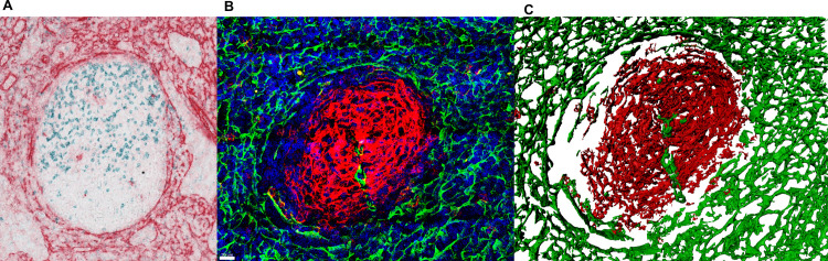
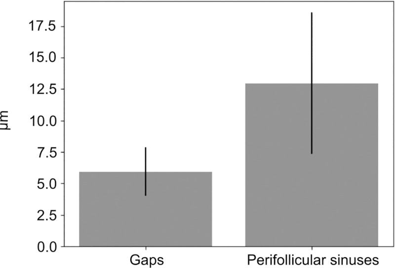
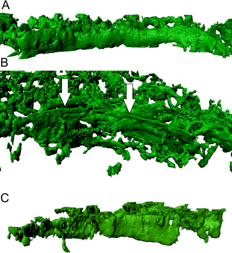
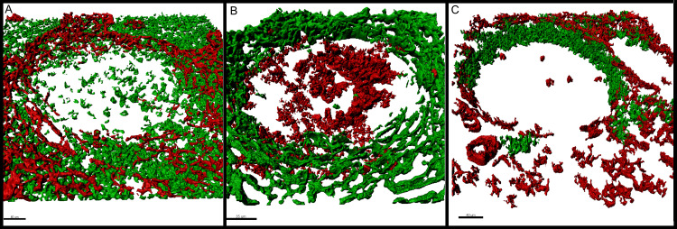
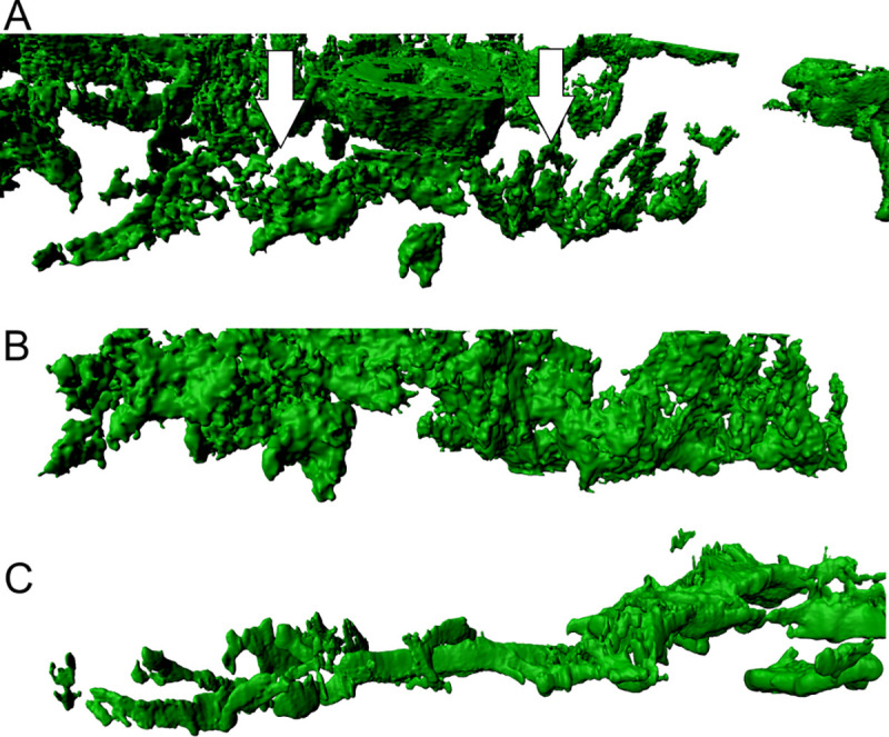
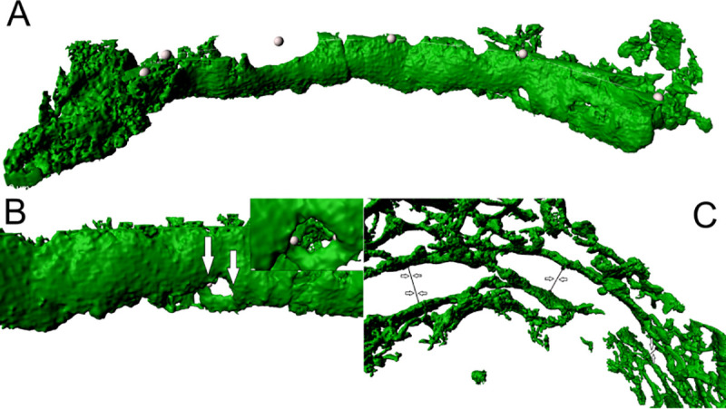
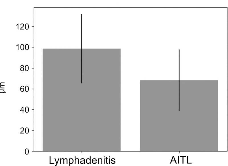
This study deals with 3D laser investigation on the border between the human lymph node T-zone and germinal centre. Only a few T-cells specific for antigen selected B-cells are allowed to enter germinal centres. This selection process is guided by sinus structures, chemokine gradients and inherent motility of the lymphoid cells. We measured gaps and wall-like structures manually, using IMARIS, a 3D image software for analysis and interpretation of microscopy datasets. In this paper, we describe alpha-actin positive and semipermeable walls and wall-like structures that may hinder T-cells and other cell types from entering germinal centres. Some clearly defined holes or gaps probably regulate lymphoid traffic between T- and B-cell areas. In lymphadenitis, the morphology of this border structure is clearly defined. However, in case of malignant lymphoma, the wall-like structure is disrupted. This has been demonstrated exemplarily in case of angioimmunoblastic T-cell lymphoma. We revealed significant differences of lengths of the wall-like structures in angioimmunoblastic T-cell lymphoma in comparison with wall-like structures in reactive tissue slices. The alterations of morphological structures lead to abnormal and less controlled T- and B-cell distributions probably preventing the immune defence against tumour cells and infectious agents by dysregulating immune homeostasis.
本研究针对人类淋巴结 T 区和生发中心之间的边界进行了 3D 激光研究。只有少数针对抗原选择 B 细胞的 T 细胞被允许进入生发中心。这个选择过程由窦结构、趋化因子梯度和淋巴细胞的固有运动指导。我们使用 IMARIS(一种用于分析和解释显微镜数据集的 3D 图像软件)手动测量间隙和壁状结构。在本文中,我们描述了可能阻碍 T 细胞和其他细胞类型进入生发中心的α-肌动蛋白阳性和半透壁状结构。一些明确界定的孔或间隙可能调节 T 细胞和 B 细胞区之间的淋巴流量。在淋巴结炎中,这种边界结构的形态清晰可见。然而,在恶性淋巴瘤的情况下,壁状结构被破坏。在血管免疫母细胞性 T 细胞淋巴瘤的病例中,我们已经证明了这一点。我们发现血管免疫母细胞性 T 细胞淋巴瘤中的壁状结构长度与反应性组织切片中的壁状结构有显著差异。形态结构的改变导致 T 细胞和 B 细胞分布异常和控制减少,可能通过失调免疫稳态来阻止对肿瘤细胞和传染性病原体的免疫防御。