• 文献检索
  • 文档翻译
  • 深度研究
  • 学术资讯
  • Suppr Zotero 插件Zotero 插件
  • 邀请有礼
  • 套餐&价格
  • 历史记录
应用&插件
Suppr Zotero 插件Zotero 插件浏览器插件Mac 客户端Windows 客户端微信小程序
定价
高级版会员购买积分包购买API积分包
服务
文献检索文档翻译深度研究API 文档MCP 服务
关于我们
关于 Suppr公司介绍联系我们用户协议隐私条款
关注我们

Suppr 超能文献

核心技术专利:CN118964589B侵权必究
粤ICP备2023148730 号-1Suppr @ 2026

文献检索

告别复杂PubMed语法,用中文像聊天一样搜索,搜遍4000万医学文献。AI智能推荐,让科研检索更轻松。

立即免费搜索

文件翻译

保留排版,准确专业,支持PDF/Word/PPT等文件格式,支持 12+语言互译。

免费翻译文档

深度研究

AI帮你快速写综述,25分钟生成高质量综述,智能提取关键信息,辅助科研写作。

立即免费体验

胸部包虫囊肿的放射学表现:肺部及肺外表现

Radiological manifestations of thoracic hydatid cysts: pulmonary and extrapulmonary findings.

作者信息

Durhan Gamze, Tan Aziz Anıl, Düzgün Selin Ardalı, Akkaya Selçuk, Arıyürek Orhan Macit

机构信息

Department of Radiology, Hacettepe University Faculty of Medicine, 06410, Ankara, Turkey.

Department of Radiology, Karadeniz Technical University Faculty of Medicine, Trabzon, Turkey.

出版信息

Insights Imaging. 2020 Nov 11;11(1):116. doi: 10.1186/s13244-020-00916-0.

DOI:10.1186/s13244-020-00916-0
PMID:33175295
原文链接:https://pmc.ncbi.nlm.nih.gov/articles/PMC7658283/
Abstract

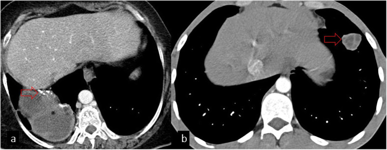
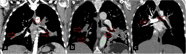
Hydatid cyst caused by the larval form of Echinococcus is a worldwide zoonosis. The lungs and liver are the most common sites involved. While the lung parenchyma is the most common site within the thorax, it may develop in any extrapulmonary region including the pleural cavity, fissures, mediastinum, heart, vascular structures, chest wall, and diaphragm. Imaging plays a pivotal role not only in the diagnosis of hydatid cyst, but also in the visualization of the extent of involvement and complications. The aim of this pictorial review was to comprehensively describe the imaging findings of thoracic hydatid cyst including pulmonary and very unusual extrapulmonary involvements. An outline is also given for the findings of complications and differential diagnosis of thoracic hydatid cyst.

摘要

由棘球绦虫幼虫形态引起的包虫囊肿是一种全球性人畜共患病。肺和肝是最常受累的部位。虽然肺实质是胸部最常见的受累部位,但它也可能发生在任何肺外区域,包括胸膜腔、叶间裂、纵隔、心脏、血管结构、胸壁和膈肌。影像学不仅在包虫囊肿的诊断中起着关键作用,而且在显示受累范围和并发症方面也很重要。本图谱综述的目的是全面描述胸部包虫囊肿的影像学表现,包括肺部及非常见的肺外受累情况。还概述了胸部包虫囊肿的并发症及鉴别诊断结果。

https://cdn.ncbi.nlm.nih.gov/pmc/blobs/b2fb/7658283/d562d9b97f79/13244_2020_916_Fig18_HTML.jpg
https://cdn.ncbi.nlm.nih.gov/pmc/blobs/b2fb/7658283/74665c50c1a4/13244_2020_916_Fig1_HTML.jpg
https://cdn.ncbi.nlm.nih.gov/pmc/blobs/b2fb/7658283/23cac1521d77/13244_2020_916_Fig2_HTML.jpg
https://cdn.ncbi.nlm.nih.gov/pmc/blobs/b2fb/7658283/377ac66638db/13244_2020_916_Fig3_HTML.jpg
https://cdn.ncbi.nlm.nih.gov/pmc/blobs/b2fb/7658283/c677415e7b6f/13244_2020_916_Fig4_HTML.jpg
https://cdn.ncbi.nlm.nih.gov/pmc/blobs/b2fb/7658283/c77be07216c2/13244_2020_916_Fig5_HTML.jpg
https://cdn.ncbi.nlm.nih.gov/pmc/blobs/b2fb/7658283/95043902f17b/13244_2020_916_Fig6_HTML.jpg
https://cdn.ncbi.nlm.nih.gov/pmc/blobs/b2fb/7658283/bb9629bbdac2/13244_2020_916_Fig7_HTML.jpg
https://cdn.ncbi.nlm.nih.gov/pmc/blobs/b2fb/7658283/4be414130ac3/13244_2020_916_Fig8_HTML.jpg
https://cdn.ncbi.nlm.nih.gov/pmc/blobs/b2fb/7658283/208dd3b6de5b/13244_2020_916_Fig9_HTML.jpg
https://cdn.ncbi.nlm.nih.gov/pmc/blobs/b2fb/7658283/84c1e60bf5a1/13244_2020_916_Fig10_HTML.jpg
https://cdn.ncbi.nlm.nih.gov/pmc/blobs/b2fb/7658283/c03f8d517406/13244_2020_916_Fig11_HTML.jpg
https://cdn.ncbi.nlm.nih.gov/pmc/blobs/b2fb/7658283/d541b7361c7a/13244_2020_916_Fig12_HTML.jpg
https://cdn.ncbi.nlm.nih.gov/pmc/blobs/b2fb/7658283/b41d166aa465/13244_2020_916_Fig13_HTML.jpg
https://cdn.ncbi.nlm.nih.gov/pmc/blobs/b2fb/7658283/5f8f185d6292/13244_2020_916_Fig14_HTML.jpg
https://cdn.ncbi.nlm.nih.gov/pmc/blobs/b2fb/7658283/70eaa8f8f941/13244_2020_916_Fig15_HTML.jpg
https://cdn.ncbi.nlm.nih.gov/pmc/blobs/b2fb/7658283/6d6fa82c714e/13244_2020_916_Fig16_HTML.jpg
https://cdn.ncbi.nlm.nih.gov/pmc/blobs/b2fb/7658283/ea364d0aa561/13244_2020_916_Fig17_HTML.jpg
https://cdn.ncbi.nlm.nih.gov/pmc/blobs/b2fb/7658283/d562d9b97f79/13244_2020_916_Fig18_HTML.jpg

相似文献

1
Radiological manifestations of thoracic hydatid cysts: pulmonary and extrapulmonary findings.胸部包虫囊肿的放射学表现:肺部及肺外表现
Insights Imaging. 2020 Nov 11;11(1):116. doi: 10.1186/s13244-020-00916-0.
2
Extrapulmonary intrathoracic hydatid cysts.肺外胸腔内包虫囊肿
Can J Surg. 2004 Apr;47(2):95-8.
3
Unusually located hydatid cysts: intrathoracic but extrapulmonary.位置异常的包虫囊肿:位于胸腔但在肺外。
Ann Thorac Surg. 1997 Aug;64(2):334-7. doi: 10.1016/S0003-4975(97)00521-3.
4
Local complications of hydatid disease involving thoracic cavity: imaging findings.累及胸腔的包虫病局部并发症:影像学表现
Eur J Radiol. 2009 Apr;70(1):49-56. doi: 10.1016/j.ejrad.2008.01.002. Epub 2008 Mar 4.
5
The evaluation of 13 patients with intrathoracic extrapulmonary hydatidosis.13例胸内肺外包虫病患者的评估。
J Int Med Res. 2005 Mar-Apr;33(2):215-21. doi: 10.1177/147323000503300209.
6
Surgical treatment and complications of thoracic hydatid disease. Report of 61 cases.胸段包虫病的外科治疗及并发症。61例报告。
Eur J Respir Dis. 1983 Apr;64(3):217-21.
7
An unusual cause of pulmonary hypertension: Mediastinal hydatid cyst.肺动脉高压的一种罕见病因:纵隔包虫囊肿。
Radiol Case Rep. 2021 Nov 1;17(1):86-90. doi: 10.1016/j.radcr.2021.09.066. eCollection 2022 Jan.
8
Do the Hydatid Cysts Have Unusual Localization and Dissemination Ways in the Chest Cavity?胸腔内的包虫囊肿是否具有不寻常的定位和传播方式?
Case Rep Surg. 2016;2016:7092494. doi: 10.1155/2016/7092494. Epub 2016 Mar 29.
9
Hydatid Disease: A Radiological Pictorial Review of a Great Neoplasms Mimicker.包虫病:一种极易被误诊为肿瘤的疾病的影像学图像综述
Diagnostics (Basel). 2023 Mar 16;13(6):1127. doi: 10.3390/diagnostics13061127.
10
Surgical approach for patients with unusually located hydatid cyst.针对位置异常的包虫囊肿患者的手术方法。
Ann Ital Chir. 2014 Jan-Feb;85(1):50-5.

引用本文的文献

1
Clinical image: multiple pulmonary, hepatic and Abdominal Wall hydatid cysts.临床影像:多发肺、肝及腹壁包虫囊肿。
Oxf Med Case Reports. 2025 Jun 27;2025(6):omaf085. doi: 10.1093/omcr/omaf085. eCollection 2025 Jun.
2
Video-Assisted Thoracoscopic Surgery (VATS) Versus Thoracotomy in the Treatment of Pulmonary Hydatid Cysts: A Randomized Controlled Trial.电视辅助胸腔镜手术(VATS)与开胸手术治疗肺包虫囊肿的随机对照试验
Health Sci Rep. 2025 Jun 11;8(6):e70668. doi: 10.1002/hsr2.70668. eCollection 2025 Jun.
3
A Rare Case of Mediastinal Hydatidosis With Cardiac and Vascular Compression.

本文引用的文献

1
A Primary Pleural Hydatid Cyst in an Unusual Location.一例罕见部位的原发性胸膜包虫囊肿
Tanaffos. 2017;16(2):166-169.
2
Computed Tomography (CT) Findings of Pulmonary Hydatid Cysts in Children and the Factors Related to Cyst Rupture.儿童肺包虫囊肿的 CT 表现及与囊肿破裂相关的因素。
Med Sci Monit. 2017 Jul 29;23:3679-3686. doi: 10.12659/msm.906163.
3
Radiological manifestations of hydatid disease and its complications.包虫病及其并发症的影像学表现。
一例罕见的伴有心脏和血管受压的纵隔包虫病
Cureus. 2025 May 4;17(5):e83483. doi: 10.7759/cureus.83483. eCollection 2025 May.
4
Pulmonary echinococcosis mimicking tuberculosis in a child from a dual-endemic region: a case report.来自双流行区的一名儿童中酷似肺结核的肺包虫病:病例报告
Front Pediatr. 2025 Apr 7;13:1562829. doi: 10.3389/fped.2025.1562829. eCollection 2025.
5
Successful off-pump resection of apical cardiac hydatid cyst: A rare case report.非体外循环下成功切除心尖部包虫囊肿:一例罕见病例报告。
Medicine (Baltimore). 2025 Mar 14;104(11):e41811. doi: 10.1097/MD.0000000000041811.
6
Massive Pulmonary Hemorrhage in a Patient With Multiple Pulmonary Cavitary Lesions: A Case Report and Literature Review.一名患有多个肺空洞性病变患者的大量肺出血:病例报告及文献综述
Cureus. 2025 Jan 21;17(1):e77787. doi: 10.7759/cureus.77787. eCollection 2025 Jan.
7
Bilateral Pulmonary Cystic Lesions with Fatal Outcome Due to Echinococcosis.因棘球蚴病导致双侧肺囊性病变并出现致命结局
Am J Trop Med Hyg. 2025 Feb 18;112(5):948-949. doi: 10.4269/ajtmh.24-0511. Print 2025 May 7.
8
A Comprehensive Analysis of Infections in Children and Adolescents: Results of a 7-Year Retrospective Study and Literature Review.儿童和青少年感染情况的综合分析:一项7年回顾性研究及文献综述的结果
Pathogens. 2025 Jan 10;14(1):53. doi: 10.3390/pathogens14010053.
9
Coexistence of kidney and lung hydatid cyst in a child: A case report.儿童肾和肺包虫囊肿并存:一例报告
Respir Med Case Rep. 2024 Nov 28;52:102138. doi: 10.1016/j.rmcr.2024.102138. eCollection 2024.
10
Non-metastatic causes of multiple pulmonary nodules.多发性肺结节的非转移性病因。
Insights Imaging. 2024 Nov 29;15(1):288. doi: 10.1186/s13244-024-01856-9.
Trop Parasitol. 2016 Jul-Dec;6(2):103-112. doi: 10.4103/2229-5070.190812.
4
Imaging in pulmonary hydatid cysts.肺包虫囊肿的影像学检查
World J Radiol. 2016 Jun 28;8(6):581-7. doi: 10.4329/wjr.v8.i6.581.
5
Multimodality imaging in diagnosis and management of alveolar echinococcosis: an update.多模态影像学在细粒棘球蚴病的诊断和治疗中的应用:最新进展。
Diagn Interv Radiol. 2016 May-Jun;22(3):247-56. doi: 10.5152/dir.2015.15456.
6
Cystic pulmonary hydatidosis.囊性肺包虫病
Lung India. 2016 Mar-Apr;33(2):179-91. doi: 10.4103/0970-2113.177449.
7
Pulmonary Embolism Originating from a Hepatic Hydatid Cyst Ruptured into the Inferior Vena Cava: CT and MRI Findings.源于破裂入下腔静脉的肝包虫囊肿的肺栓塞:CT和MRI表现
Case Rep Radiol. 2016;2016:3589812. doi: 10.1155/2016/3589812. Epub 2016 Jan 21.
8
Management of complicated hydatid cyst of the thorax.胸部复杂包虫囊肿的管理
ANZ J Surg. 2007 Sep;77(9):752-7. doi: 10.1111/j.1445-2197.2007.04217.x.
9
Comparison of pulmonary hydatid cysts in children and adults.儿童与成人肺包虫囊肿的比较。
ANZ J Surg. 2004 Oct;74(10):885-9. doi: 10.1111/j.1445-1433.2004.03022.x.
10
Extrapulmonary intrathoracic hydatid cysts.肺外胸腔内包虫囊肿
Can J Surg. 2004 Apr;47(2):95-8.