British Heart Foundation Centre for Cardiovascular Science, University of Edinburgh, Chancellor's Building, 49 Little France, Edinburgh, EH16 4TJ, UK.
J Nucl Cardiol. 2021 Apr;28(2):481-491. doi: 10.1007/s12350-020-02411-x. Epub 2020 Nov 11.
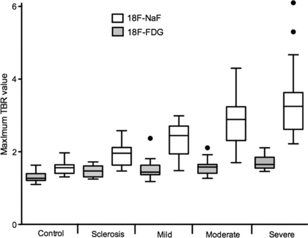
Calcific aortic valve disease is the most common valvular disease and confers significant morbidity and mortality. There are currently no medical therapies that successfully halt or reverse the disease progression, making surgical replacement the only treatment currently available. The majority of patients will receive a bioprosthetic valve, which themselves are prone to degeneration and may also need replaced, adding to the already substantial healthcare burden of aortic stenosis. Echocardiography and computed tomography can identify late-stage manifestations of the disease process affecting native and bioprosthetic aortic valves but cannot detect or quantify early molecular changes. F-fluoride positron emission tomography, on the other hand, can non-invasively and sensitively assess disease activity in the valves. The current review outlines the pivotal role this novel molecular imaging technique has played in improving our understanding of native and bioprosthetic aortic valve disease, as well as providing insights into its feasibility as an important future research and clinical tool.
钙化性主动脉瓣疾病是最常见的瓣膜病,会导致显著的发病率和死亡率。目前尚无成功阻止或逆转疾病进展的医学治疗方法,因此手术置换是目前唯一可用的治疗方法。大多数患者将接受生物瓣,而生物瓣本身容易退化,也可能需要更换,这增加了主动脉瓣狭窄已经很大的医疗保健负担。超声心动图和计算机断层扫描可以识别影响原生和生物瓣的疾病过程的晚期表现,但不能检测或量化早期分子变化。另一方面,氟-18 正电子发射断层扫描可以非侵入性且敏感地评估瓣膜中的疾病活动。目前的综述概述了这项新的分子成像技术在提高我们对原生和生物瓣主动脉瓣疾病的认识方面所起的关键作用,并探讨了它作为一种重要的未来研究和临床工具的可行性。