British Heart Foundation Centre for Cardiovascular Science, University of Edinburgh, Edinburgh, Scotland, United Kingdom.
National Heart and Lung Institute, Imperial College London, London, United Kingdom.
JACC Cardiovasc Imaging. 2019 Jan;12(1):185-197. doi: 10.1016/j.jcmg.2018.10.023.
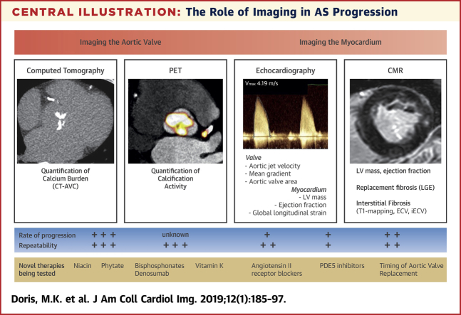
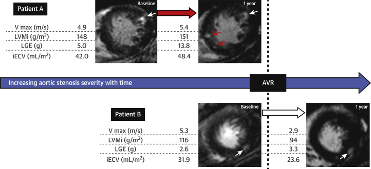
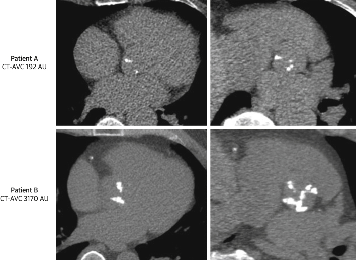
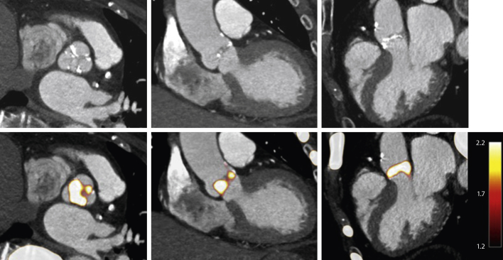
Aortic stenosis represents a growing health care burden in high-income countries. Currently, the only definitive treatment is surgical or transcatheter valve intervention at the end stages of disease. As the understanding of the underlying pathophysiology evolves, many promising therapies are being investigated. These seek to both slow disease progression in the valve and delay the transition from hypertrophy to heart failure in the myocardium, with the ultimate aim of avoiding the need for valve replacement in the elderly patients afflicted by this condition. Noninvasive imaging has played a pivotal role in enhancing our understanding of the complex pathophysiology underlying aortic stenosis, as well as disease progression in both the valve and myocardium. In this review, the authors discuss the means by which contemporary imaging may be used to assess disease progression and how these approaches may be utilized, both in clinical practice and research trials exploring the clinical efficacy of novel therapies.
主动脉瓣狭窄是高收入国家日益严重的健康问题。目前,该病的唯一根治方法是在疾病晚期进行手术或经导管瓣膜介入治疗。随着对潜在病理生理学的认识不断发展,许多有前途的治疗方法正在被研究。这些治疗方法旨在减缓瓣膜疾病的进展,并延缓心肌从肥厚到心力衰竭的转变,最终目的是避免老年患者需要更换瓣膜。非侵入性成像在增强我们对主动脉瓣狭窄基础复杂病理生理学以及瓣膜和心肌疾病进展的理解方面发挥了关键作用。在这篇综述中,作者讨论了如何使用现代成像方法来评估疾病进展,以及如何在临床实践和研究试验中利用这些方法来探索新型治疗方法的临床疗效。