Dipartimento di Medicina Veterinaria, Università degli Studi di Sassari, Sassari, Italy.
Facoltà di Medicina e Chirurgia, Università degli Studi di Sassari, Sassari, Italy.
Parasit Vectors. 2020 Nov 11;13(1):568. doi: 10.1186/s13071-020-04439-x.
Cysticercosis caused by cysticercus tenuicollis is a metacestode infection that affects several species of ungulates. It is caused by the larval stage of Taenia hydatigena, an intestinal tapeworm in dogs and wild canids. In the intermediate host, the mature cysticerci are usually found in the omentum, mesentery, and peritoneum, and less frequently in the pleura and pericardium. The migrating larvae can be found mostly in the liver parenchyma causing traumatic hepatitis in young animals. Most infections are chronic and asymptomatic, and are diagnosed at the abattoir. The acute form of infection is unusual in sheep and reports of death in lambs are rare.
In March 2018, fifteen female lambs presented anorexia, weakness, lethargy, and death secondary to acute visceral cysticercosis. Twelve of them underwent hepatic ultrasonography. Examinations were performed on standing or left lateral recumbent animals.
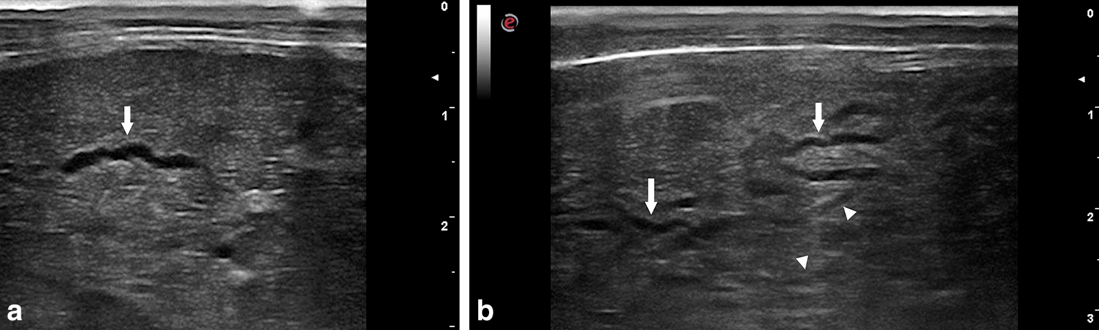
Livers of affected animals presented rounded margins and a thickened, irregular and hyperechoic surface. Hepatic parenchyma appeared to be wholly or partially affected by lesions characterized by heterogeneous areas crossed by numerous, irregular, anechoic tracts ranging from 1 to 2 cm in length and 0.1 to 0.2 cm in width. Superficial and intraparenchymal cystic structures were also visualized. The presence of lesions was confirmed by anatomopathological examination, and T. hydatigena cysticerci was identified by morphological and molecular characterization of isolates.
Our results highlighted that hepatic ultrasonography is effective for an intra-vitam diagnosis of acute cysticercosis in lambs.
由细粒棘球蚴引起的囊尾蚴病是一种囊蚴感染,影响多种有蹄类动物。它是由犬和野生犬科动物肠道带绦虫细粒棘球绦虫的幼虫阶段引起的。在中间宿主中,成熟的囊尾蚴通常在大网膜、肠系膜和腹膜中发现,在胸膜和心包中较少见。迁移的幼虫主要在肝实质中发现,导致幼小动物外伤性肝炎。大多数感染是慢性和无症状的,并在屠宰场诊断。绵羊的急性感染形式不常见,羔羊死亡的报道很少。
2018 年 3 月,15 只母羊出现厌食、虚弱、昏睡和死于急性内脏囊尾蚴病。其中 12 只进行了肝超声检查。在站立或左侧横卧的动物身上进行检查。
受影响动物的肝脏呈现圆形边缘和增厚、不规则和高回声表面。肝实质似乎全部或部分受到病变的影响,病变特征为不均匀区域,由许多不规则的无回声道交叉,长度为 1 至 2 厘米,宽度为 0.1 至 0.2 厘米。还观察到浅表和肝内囊性结构。解剖病理学检查证实了病变的存在,并通过对分离物的形态学和分子特征鉴定出细粒棘球绦虫囊尾蚴。
我们的结果强调了肝超声检查对于羔羊急性囊尾蚴病的活体诊断是有效的。