• 文献检索
  • 文档翻译
  • 深度研究
  • 学术资讯
  • Suppr Zotero 插件Zotero 插件
  • 邀请有礼
  • 套餐&价格
  • 历史记录
应用&插件
Suppr Zotero 插件Zotero 插件浏览器插件Mac 客户端Windows 客户端微信小程序
定价
高级版会员购买积分包购买API积分包
服务
文献检索文档翻译深度研究API 文档MCP 服务
关于我们
关于 Suppr公司介绍联系我们用户协议隐私条款
关注我们

Suppr 超能文献

核心技术专利:CN118964589B侵权必究
粤ICP备2023148730 号-1Suppr @ 2026

文献检索

告别复杂PubMed语法,用中文像聊天一样搜索,搜遍4000万医学文献。AI智能推荐,让科研检索更轻松。

立即免费搜索

文件翻译

保留排版,准确专业,支持PDF/Word/PPT等文件格式,支持 12+语言互译。

免费翻译文档

深度研究

AI帮你快速写综述,25分钟生成高质量综述,智能提取关键信息,辅助科研写作。

立即免费体验

单细胞分析揭示严重 COVID-19 感染中的 T 细胞过度激活和瘫痪。

T-Cell Hyperactivation and Paralysis in Severe COVID-19 Infection Revealed by Single-Cell Analysis.

机构信息

Department of Life Sciences, Imperial College London, London, United Kingdom.

Instituto Gulbenkian de Ciência, Oeiras, Portugal.

出版信息

Front Immunol. 2020 Oct 8;11:589380. doi: 10.3389/fimmu.2020.589380. eCollection 2020.

DOI:10.3389/fimmu.2020.589380
PMID:33178221
原文链接:https://pmc.ncbi.nlm.nih.gov/articles/PMC7596772/
Abstract

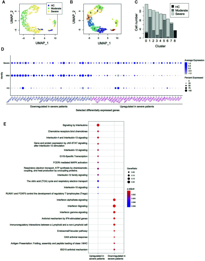
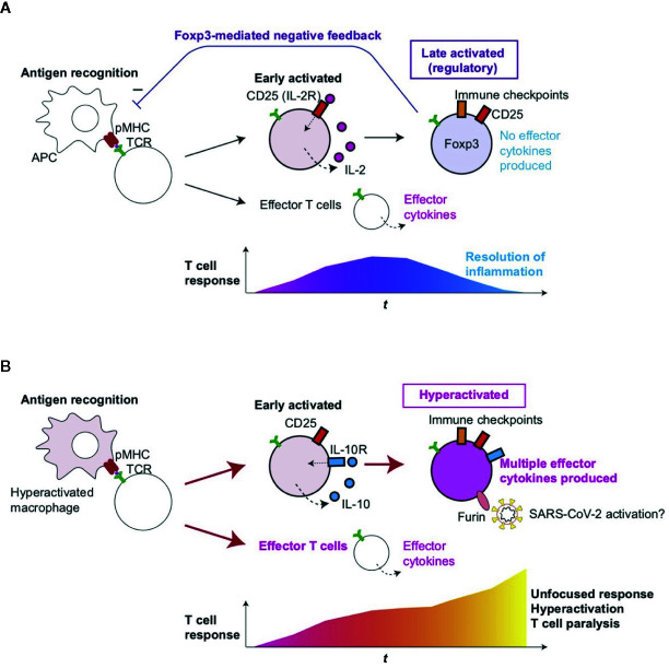
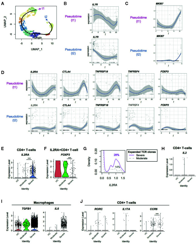
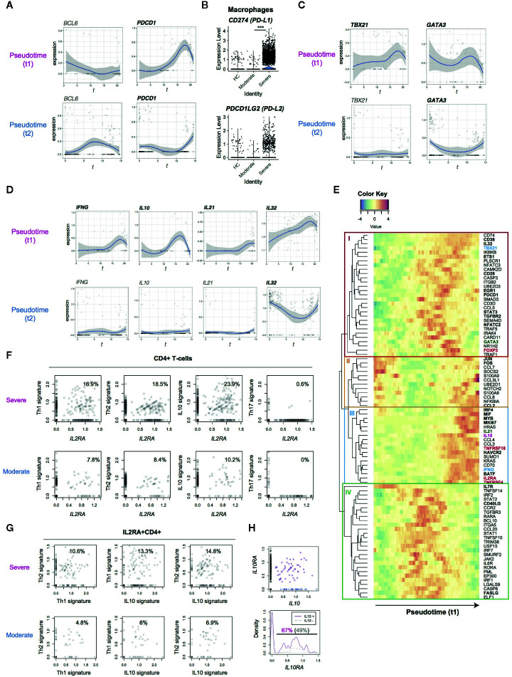
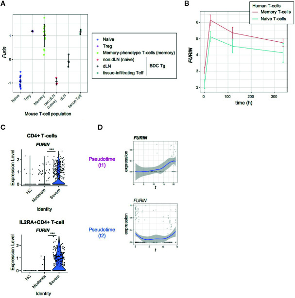
Severe COVID-19 patients show various immunological abnormalities including T-cell reduction and cytokine release syndrome, which can be fatal and is a major concern of the pandemic. However, it is poorly understood how T-cell dysregulation can contribute to the pathogenesis of severe COVID-19. Here we show single cell-level mechanisms for T-cell dysregulation in severe COVID-19, demonstrating new pathogenetic mechanisms of T-cell activation and differentiation underlying severe COVID-19. By sorting CD4+ T-cells from a single cell RNA-seq dataset, we found that CD4+ T-cells were highly activated and showed unique differentiation pathways in the lung of severe COVID-19 patients. Notably, those T-cells in severe COVID-19 patients highly expressed immunoregulatory receptors and CD25, whilst repressing the expression of FOXP3. Furthermore, we show that CD25 hyperactivated T-cells differentiate into multiple helper T-cell lineages, showing multifaceted effector T-cells with Th1 and Th2 characteristics. Lastly, we show that CD25-expressing hyperactivated T-cells produce the protease Furin, which facilitates the viral entry of SARS-CoV-2. Collectively, CD4 T-cells from severe COVID-19 patients are hyperactivated and FOXP3-mediated negative feedback mechanisms are impaired in the lung, which may promote immunopathology. Therefore, our study proposes a new model of T-cell hyperactivation and paralysis that drives immunopathology in severe COVID-19.

摘要

严重 COVID-19 患者表现出各种免疫异常,包括 T 细胞减少和细胞因子释放综合征,这可能是致命的,也是大流行的主要关注点。然而,人们对 T 细胞失调如何导致严重 COVID-19 的发病机制知之甚少。在这里,我们展示了严重 COVID-19 中 T 细胞失调的单细胞水平机制,证明了严重 COVID-19 中 T 细胞激活和分化的新发病机制。通过从单细胞 RNA-seq 数据集分选 CD4+ T 细胞,我们发现 CD4+ T 细胞在严重 COVID-19 患者的肺部高度激活,并表现出独特的分化途径。值得注意的是,严重 COVID-19 患者中的这些 T 细胞高度表达免疫调节受体和 CD25,同时抑制 FOXP3 的表达。此外,我们表明 CD25 过度激活的 T 细胞分化为多个辅助性 T 细胞谱系,表现出具有 Th1 和 Th2 特征的多方面效应 T 细胞。最后,我们表明表达 CD25 的过度激活的 T 细胞产生蛋白酶 Furin,这促进了 SARS-CoV-2 的病毒进入。总之,严重 COVID-19 患者的 CD4 T 细胞过度激活,肺中 FOXP3 介导的负反馈机制受损,这可能促进免疫病理学。因此,我们的研究提出了一种新的 T 细胞过度激活和麻痹模型,该模型驱动严重 COVID-19 中的免疫病理学。

https://cdn.ncbi.nlm.nih.gov/pmc/blobs/e12e/7596772/35cbca58d635/fimmu-11-589380-g005.jpg
https://cdn.ncbi.nlm.nih.gov/pmc/blobs/e12e/7596772/164abbfcfbf9/fimmu-11-589380-g001.jpg
https://cdn.ncbi.nlm.nih.gov/pmc/blobs/e12e/7596772/32517b862b07/fimmu-11-589380-g002.jpg
https://cdn.ncbi.nlm.nih.gov/pmc/blobs/e12e/7596772/2d2bd3b11a44/fimmu-11-589380-g003.jpg
https://cdn.ncbi.nlm.nih.gov/pmc/blobs/e12e/7596772/eb6051219973/fimmu-11-589380-g004.jpg
https://cdn.ncbi.nlm.nih.gov/pmc/blobs/e12e/7596772/35cbca58d635/fimmu-11-589380-g005.jpg

相似文献

1
T-Cell Hyperactivation and Paralysis in Severe COVID-19 Infection Revealed by Single-Cell Analysis.单细胞分析揭示严重 COVID-19 感染中的 T 细胞过度激活和瘫痪。
Front Immunol. 2020 Oct 8;11:589380. doi: 10.3389/fimmu.2020.589380. eCollection 2020.
2
T-cell dysregulation in COVID-19.COVID-19 中的 T 细胞失调。
Biochem Biophys Res Commun. 2021 Jan 29;538:204-210. doi: 10.1016/j.bbrc.2020.10.079. Epub 2020 Nov 7.
3
FoxP3+ CD25+ CD8+ T-cell induction during primary simian immunodeficiency virus infection in cynomolgus macaques correlates with low CD4+ T-cell activation and high viral load.食蟹猴原发性猴免疫缺陷病毒感染期间FoxP3+ CD25+ CD8+ T细胞的诱导与低水平CD4+ T细胞活化及高病毒载量相关。
J Virol. 2007 Dec;81(24):13444-55. doi: 10.1128/JVI.01466-07. Epub 2007 Sep 26.
4
Imbalance of Regulatory and Cytotoxic SARS-CoV-2-Reactive CD4 T Cells in COVID-19.COVID-19 中 SARS-CoV-2 反应性调节性和细胞毒性 CD4 T 细胞的失衡。
Cell. 2020 Nov 25;183(5):1340-1353.e16. doi: 10.1016/j.cell.2020.10.001. Epub 2020 Oct 5.
5
Tracking Regulatory T Cell Development in the Thymus Using Single-Cell RNA Sequencing/TCR Sequencing.使用单细胞 RNA 测序/TCR 测序追踪胸腺中调节性 T 细胞的发育。
J Immunol. 2022 Oct 1;209(7):1300-1313. doi: 10.4049/jimmunol.2200089. Epub 2022 Aug 29.
6
Plasmodium falciparum-mediated induction of human CD25Foxp3 CD4 T cells is independent of direct TCR stimulation and requires IL-2, IL-10 and TGFbeta.恶性疟原虫介导的人类CD25Foxp3 CD4 T细胞诱导不依赖于直接的TCR刺激,且需要白细胞介素-2、白细胞介素-10和转化生长因子β。
PLoS Pathog. 2009 Aug;5(8):e1000543. doi: 10.1371/journal.ppat.1000543. Epub 2009 Aug 14.
7
Foxp3+ CD4+ regulatory T cells control dendritic cells in inducing antigen-specific immunity to emerging SARS-CoV-2 antigens.Foxp3+ CD4+ 调节性 T 细胞控制树突状细胞诱导针对新型 SARS-CoV-2 抗原的抗原特异性免疫。
PLoS Pathog. 2021 Dec 9;17(12):e1010085. doi: 10.1371/journal.ppat.1010085. eCollection 2021 Dec.
8
Tolerogenic dendritic cells induce CD4+CD25hiFoxp3+ regulatory T cell differentiation from CD4+CD25-/loFoxp3- effector T cells.耐受性树突状细胞诱导 CD4+CD25-/loFoxp3-效应 T 细胞向 CD4+CD25hiFoxp3+调节性 T 细胞分化。
J Immunol. 2010 Nov 1;185(9):5003-10. doi: 10.4049/jimmunol.0903446. Epub 2010 Sep 24.
9
The calcineurin inhibitor tacrolimus allows the induction of functional CD4CD25 regulatory T cells by rabbit anti-thymocyte globulins.钙调磷酸酶抑制剂他克莫司通过兔抗胸腺球蛋白诱导功能性 CD4CD25 调节性 T 细胞。
Clin Exp Immunol. 2010 Aug;161(2):364-77. doi: 10.1111/j.1365-2249.2010.04183.x. Epub 2010 May 28.
10
Stimulation of α7 nicotinic acetylcholine receptor by nicotine increases suppressive capacity of naturally occurring CD4+CD25+ regulatory T cells in mice in vitro.尼古丁刺激 α7 烟碱型乙酰胆碱受体可增加体外培养的小鼠天然存在的 CD4+CD25+调节性 T 细胞的抑制能力。
J Pharmacol Exp Ther. 2010 Dec;335(3):553-61. doi: 10.1124/jpet.110.169961. Epub 2010 Sep 15.

引用本文的文献

1
The Multifaceted Role of Regulatory T Cells in Sepsis: Mechanisms, Heterogeneity, and Pathogen-Tailored Therapies.调节性T细胞在脓毒症中的多方面作用:机制、异质性及病原体针对性疗法
Int J Mol Sci. 2025 Aug 1;26(15):7436. doi: 10.3390/ijms26157436.
2
Immune transcriptomic analysis on COVID-19 patients with varying clinical presentations identifies severity markers.对具有不同临床表现的新冠肺炎患者进行免疫转录组分析,确定了疾病严重程度标志物。
Sci Rep. 2025 Jul 2;15(1):23416. doi: 10.1038/s41598-025-04837-0.
3
Aging shapes infection profiles of influenza A virus and SARS-CoV-2 in human precision-cut lung slices.

本文引用的文献

1
Immune mechanisms of pulmonary intravascular coagulopathy in COVID-19 pneumonia.新型冠状病毒肺炎肺血管内凝血病变的免疫机制
Lancet Rheumatol. 2020 Jul;2(7):e437-e445. doi: 10.1016/S2665-9913(20)30121-1. Epub 2020 May 7.
2
Reduction and Functional Exhaustion of T Cells in Patients With Coronavirus Disease 2019 (COVID-19).新型冠状病毒病(COVID-19)患者 T 细胞减少和功能耗竭。
Front Immunol. 2020 May 1;11:827. doi: 10.3389/fimmu.2020.00827. eCollection 2020.
3
Single-cell landscape of bronchoalveolar immune cells in patients with COVID-19.
衰老塑造了甲型流感病毒和新冠病毒在人精密切割肺切片中的感染特征。
Respir Res. 2025 Mar 24;26(1):112. doi: 10.1186/s12931-025-03190-0.
4
Combined PARP14 inhibition and PD-1 blockade promotes cytotoxic T cell quiescence and modulates macrophage polarization in relapsed melanoma.PARP14抑制与PD-1阻断联合应用可促进复发性黑色素瘤中细胞毒性T细胞的静止,并调节巨噬细胞极化。
J Immunother Cancer. 2025 Jan 27;13(1):e010683. doi: 10.1136/jitc-2024-010683.
5
A multidimensional toolkit for elucidating temporal trajectories in cell development in vivo.一种用于阐明体内细胞发育时间轨迹的多维工具包。
Development. 2024 Dec 15;151(24). doi: 10.1242/dev.204255. Epub 2024 Dec 18.
6
Proteomic signatures of vaccine-induced and breakthrough infection-induced host responses to SARS-CoV-2.疫苗诱导和突破性感染诱导的宿主对SARS-CoV-2反应的蛋白质组学特征
Vaccine. 2025 Jan 1;43(Pt 1):126484. doi: 10.1016/j.vaccine.2024.126484. Epub 2024 Nov 8.
7
T Regulatory Cell Subsets Do Not Restore for One Year After Acute COVID-19.急性 COVID-19 后一年,调节性 T 细胞亚群未恢复。
Int J Mol Sci. 2024 Nov 1;25(21):11759. doi: 10.3390/ijms252111759.
8
scParser: sparse representation learning for scalable single-cell RNA sequencing data analysis.scParser:用于可扩展单细胞 RNA 测序数据分析的稀疏表示学习。
Genome Biol. 2024 Aug 16;25(1):223. doi: 10.1186/s13059-024-03345-0.
9
COVID-19: from immune response to clinical intervention.新型冠状病毒肺炎:从免疫反应到临床干预
Precis Clin Med. 2024 Jul 18;7(3):pbae015. doi: 10.1093/pcmedi/pbae015. eCollection 2024 Sep.
10
SARS-CoV-2: pathogenesis, therapeutics, variants, and vaccines.严重急性呼吸综合征冠状病毒2:发病机制、治疗方法、变体和疫苗。
Front Microbiol. 2024 Jun 13;15:1334152. doi: 10.3389/fmicb.2024.1334152. eCollection 2024.
COVID-19 患者支气管肺泡免疫细胞的单细胞景观。
Nat Med. 2020 Jun;26(6):842-844. doi: 10.1038/s41591-020-0901-9. Epub 2020 May 12.
4
A Multibasic Cleavage Site in the Spike Protein of SARS-CoV-2 Is Essential for Infection of Human Lung Cells.SARS-CoV-2 刺突蛋白中的多碱性裂解位点对于感染人肺细胞至关重要。
Mol Cell. 2020 May 21;78(4):779-784.e5. doi: 10.1016/j.molcel.2020.04.022. Epub 2020 May 1.
5
SARS-CoV-2 entry factors are highly expressed in nasal epithelial cells together with innate immune genes.严重急性呼吸综合征冠状病毒2(SARS-CoV-2)进入因子与固有免疫基因一起在鼻上皮细胞中高表达。
Nat Med. 2020 May;26(5):681-687. doi: 10.1038/s41591-020-0868-6. Epub 2020 Apr 23.
6
Letter to the Editor: Obesity as a risk factor for greater severity of COVID-19 in patients with metabolic associated fatty liver disease.致编辑的信:肥胖作为代谢相关脂肪性肝病患者感染 COVID-19 时病情更严重的风险因素。
Metabolism. 2020 Jul;108:154244. doi: 10.1016/j.metabol.2020.154244. Epub 2020 Apr 19.
7
Clinical and immunological features of severe and moderate coronavirus disease 2019.新型冠状病毒病 2019 重症和中度患者的临床和免疫学特征。
J Clin Invest. 2020 May 1;130(5):2620-2629. doi: 10.1172/JCI137244.
8
Risk Factors Associated With Acute Respiratory Distress Syndrome and Death in Patients With Coronavirus Disease 2019 Pneumonia in Wuhan, China.中国武汉 2019 年冠状病毒病肺炎患者急性呼吸窘迫综合征和死亡的相关危险因素。
JAMA Intern Med. 2020 Jul 1;180(7):934-943. doi: 10.1001/jamainternmed.2020.0994.
9
SARS-CoV-2 Cell Entry Depends on ACE2 and TMPRSS2 and Is Blocked by a Clinically Proven Protease Inhibitor.严重急性呼吸综合征冠状病毒 2 型(SARS-CoV-2)进入细胞依赖于 ACE2 和 TMPRSS2,可被一种临床验证的蛋白酶抑制剂所阻断。
Cell. 2020 Apr 16;181(2):271-280.e8. doi: 10.1016/j.cell.2020.02.052. Epub 2020 Mar 5.
10
The spike glycoprotein of the new coronavirus 2019-nCoV contains a furin-like cleavage site absent in CoV of the same clade.新型冠状病毒 2019-nCoV 的刺突糖蛋白含有弗林蛋白酶样裂解位点,而同一进化枝的 CoV 中则没有。
Antiviral Res. 2020 Apr;176:104742. doi: 10.1016/j.antiviral.2020.104742. Epub 2020 Feb 10.