Darche Fabrice F, Rivinius Rasmus, Rahm Ann-Kathrin, Köllensperger Eva, Leimer Uwe, Germann Günter, Reiss Miriam, Koenen Michael, Katus Hugo A, Thomas Dierk, Schweizer Patrick A
Department of Cardiology, Medical University Hospital Heidelberg, Heidelberg D-69120, Germany.
Department of Plastic Surgery, ETHIANUM Klinik Heidelberg, Heidelberg D-69115, Germany.
World J Stem Cells. 2020 Oct 26;12(10):1133-1151. doi: 10.4252/wjsc.v12.i10.1133.
Mesenchymal stem cells (MSC) modified by gene transfer to express cardiac pacemaker channels such as HCN2 or HCN4 were shown to elicit pacemaker function after intracardiac transplantation in experimental animal models. Human MSC derived from adipose tissue (haMSC) differentiate into cells with pacemaker properties , but little is known about their behavior after intracardiac transplantation.
To investigate whether haMSC elicit biological pacemaker function after transplantation into pig hearts.
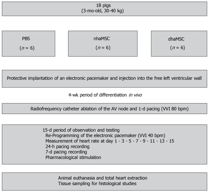
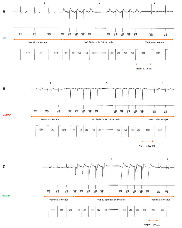
haMSC under native conditions (nhaMSC) or after pre-conditioning by medium differentiation (dhaMSC) ( = 6 pigs each, 5 × 10 cells/animal) were injected into the porcine left ventricular free wall. Animals receiving PBS injection served as controls ( = 6). Four weeks later, total atrioventricular (AV)-block was induced by radiofrequency catheter ablation, and electronic pacemaker devices were implanted for backup stimulation and heart rate monitoring. Ventricular rate and rhythm of pigs were evaluated during a follow-up of 15 d post ablation by 12-lead-ECG with heart rate assessment, 24-h continuous rate monitoring recorded by electronic pacemaker, assessment of escape recovery time, and pharmacological challenge to address catecholaminergic rate response. Finally, hearts were analyzed by histological and immunohistochemical investigations.
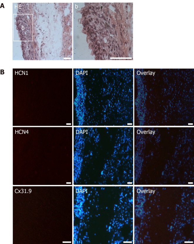
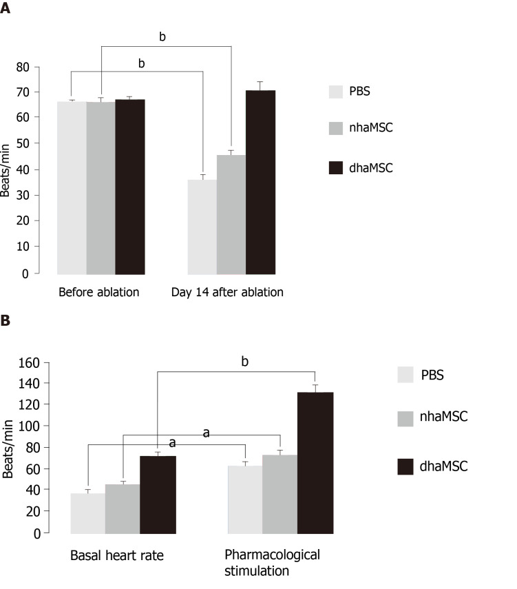
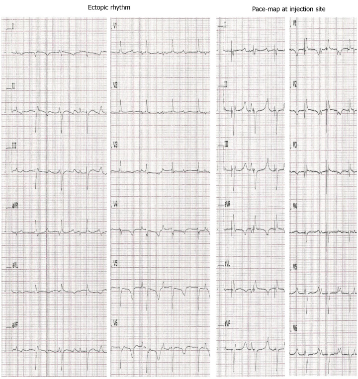
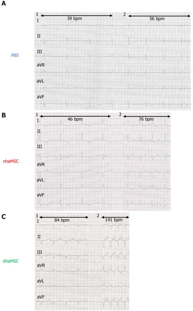
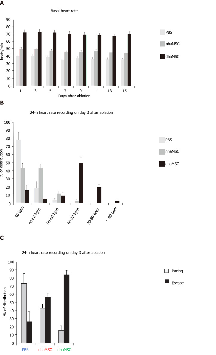
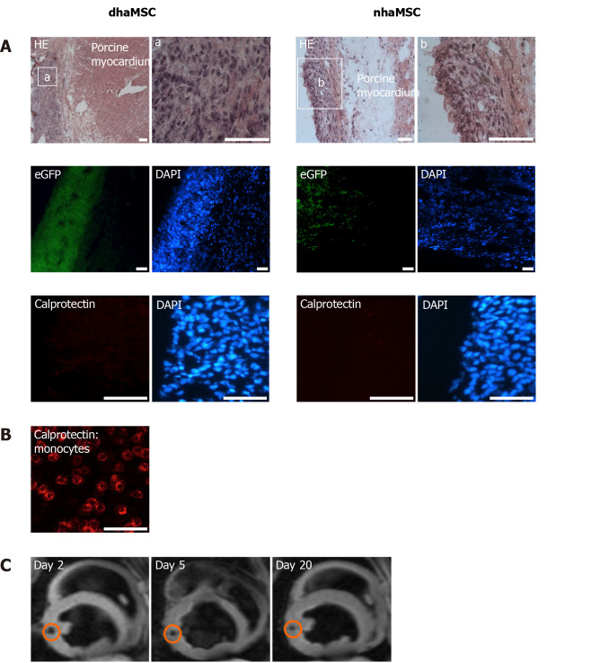
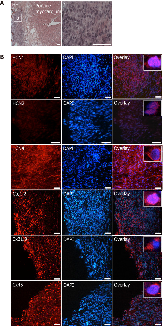
transplantation of dhaMSC into the left ventricular free wall of pigs elicited spontaneous and regular rhythms that were pace-mapped to ventricular injection sites (mean heart rate 72.2 ± 3.6 bpm; 6) after experimental total AV block. Ventricular rhythms were stably detected over a 15-d period and were sensitive to catecholaminergic stimulation (mean maximum heart rate 131.0 ± 6.2 bpm; 6; 0.001). Pigs, which received nhaMSC or PBS presented significantly lower ventricular rates (mean heart rates 47.2 ± 2.5 bpm and 37.4 ± 3.2 bpm, respectively; 6 each; < 0.001) and exhibited little sensitivity towards catecholaminergic stimulation (mean maximum heart rates 76.4 ± 3.1 bpm and 60.5 ± 3.1 bpm, respectively; 6 each; < 0.05). Histological and immunohistochemical evaluation of hearts treated with dhaMSC revealed local clusters of transplanted cells at the injection sites that lacked macrophage or lymphocyte infiltrations or tumor formation. Intense fluorescence signals at these sites indicated membrane expression of HCN4 and other pacemaker-specific proteins involved in cardiac automaticity and impulse propagation.
dhaMSC transplanted into pig left ventricles sustainably induced rate-responsive ventricular pacemaker activity after engraftment for four weeks. The data suggest that pre-conditioned MSC may further differentiate along a pacemaker-related lineage after myocardial integration and may establish superior pacemaker properties .
在实验动物模型中,经基因转移修饰以表达心脏起搏器通道(如HCN2或HCN4)的间充质干细胞(MSC)在心脏内移植后可引发起搏器功能。源自脂肪组织的人MSC(haMSC)可分化为具有起搏器特性的细胞,但关于其心脏内移植后的行为知之甚少。
研究haMSC移植到猪心脏后是否能引发生物起搏器功能。
将处于天然状态的haMSC(nhaMSC)或经培养基分化预处理后的haMSC(dhaMSC)(每组6头猪,5×10⁶个细胞/只动物)注射到猪左心室游离壁。接受PBS注射的动物作为对照(每组6只)。4周后,通过射频导管消融诱导完全房室(AV)阻滞,并植入电子起搏器装置用于备用刺激和心率监测。在消融后15天的随访期间,通过12导联心电图评估猪的心室率和节律,并进行心率评估,通过电子起搏器记录24小时连续心率监测,评估逸搏恢复时间,并进行药理学激发以评估儿茶酚胺能心率反应。最后,通过组织学和免疫组织化学研究分析心脏。
将dhaMSC移植到猪左心室游离壁后,在实验性完全AV阻滞诱导后引发了自发且规则的节律,这些节律通过起搏标测定位于心室注射部位(平均心率72.2±3.6次/分钟;n = 6)。在15天的时间内稳定检测到心室节律,并且对儿茶酚胺能刺激敏感(平均最大心率131.0±6.2次/分钟;n = 6;P < 0.001)。接受nhaMSC或PBS的猪心室率明显较低(平均心率分别为47.2±2.5次/分钟和37.4±3.2次/分钟;每组n = 6;P < 0.001),并且对儿茶酚胺能刺激的敏感性较低(平均最大心率分别为76.4±3.1次/分钟和60.5±3.1次/分钟;每组n = 6;P < 0.05)。对接受dhaMSC治疗的心脏进行组织学和免疫组织化学评估发现,注射部位有局部移植细胞簇,缺乏巨噬细胞或淋巴细胞浸润或肿瘤形成。这些部位强烈的荧光信号表明HCN4和其他参与心脏自律性和冲动传播的起搏器特异性蛋白的膜表达。
移植到猪左心室的dhaMSC在植入4周后可持续诱导心率反应性心室起搏器活动。数据表明,预处理的MSC在心肌整合后可能沿起搏器相关谱系进一步分化,并可能建立更优越的起搏器特性。