Mannino Giuliana, Gennuso Florinda, Giurdanella Giovanni, Conti Federica, Drago Filippo, Salomone Salvatore, Furno Debora Lo, Bucolo Claudio, Giuffrida Rosario
Physiology Section, Department of Biomedical and Biotechnological Sciences, University of Catania, Catania 95123, Italy.
Pharmacology Section, Department of Biomedical and Biotechnological Sciences, School of Medicine, University of Catania, Catania 95123, Italy.
World J Stem Cells. 2020 Oct 26;12(10):1152-1170. doi: 10.4252/wjsc.v12.i10.1152.
Adipose-derived mesenchymal stem cells (ASCs) are characterized by long-term self-renewal and a high proliferation rate. Under adequate conditions, they may differentiate into cells belonging to mesodermal, endodermal or ectodermal lineages. Pericytes support endothelial cells and play an important role in stabilizing the vessel wall at the microcirculation level. The loss of pericytes, as occurs in diabetic retinopathy, results in a breakdown of the blood-retina barrier (BRB) and infiltration of inflammatory cells. In this context, the use of pericyte-like differentiated ASCs may represent a valuable therapeutic strategy for restoring BRB damage.
To test strategies to obtain pericyte-like differentiation of human ASCs (hASCs).
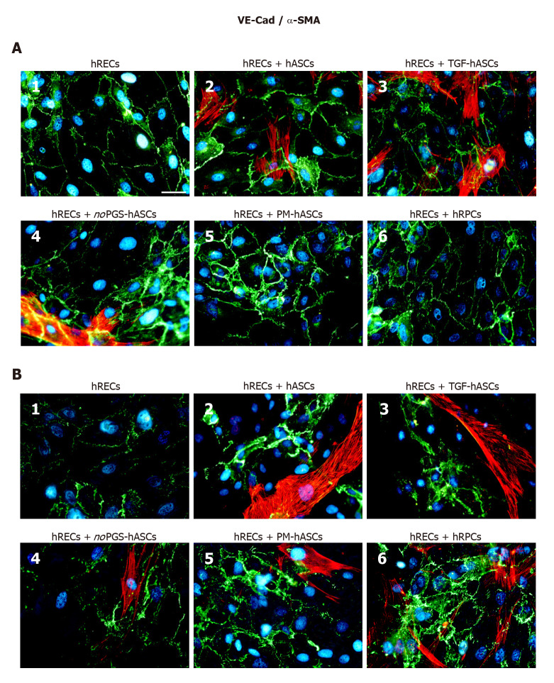
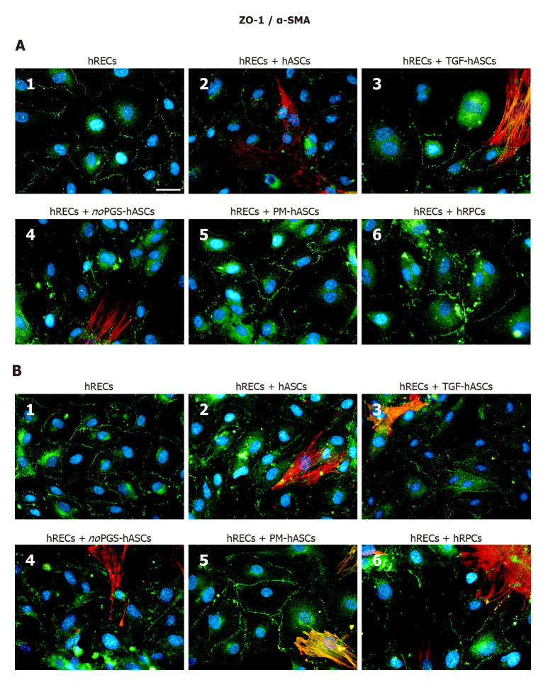
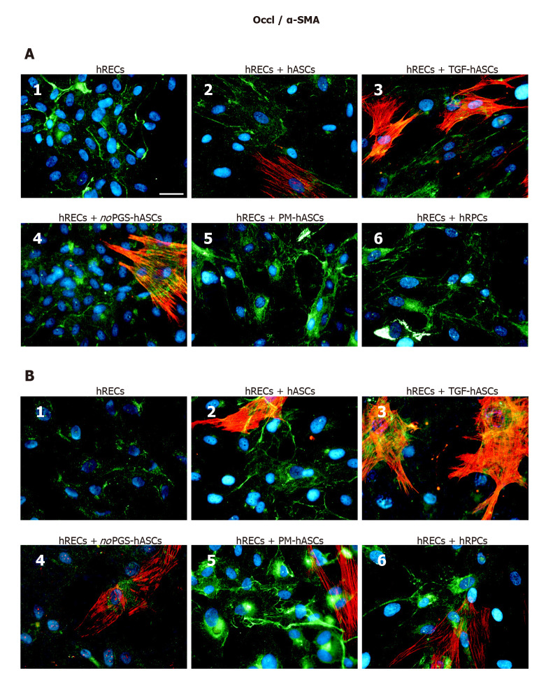
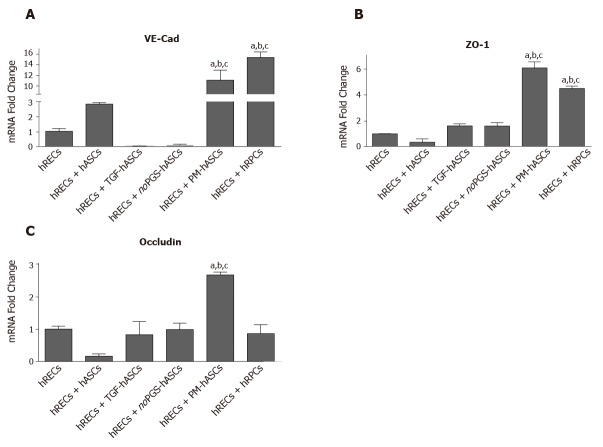
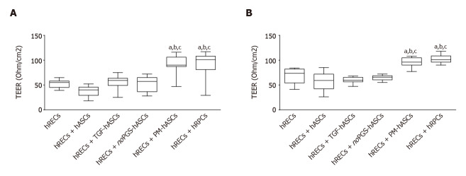
Different culture conditions were tested: hASCs cultured in a basal medium supplemented with transforming growth factor β1; and hASCs cultured in a specific pericyte medium (PM-hASCs). In a further sample, pericyte growth supplement was omitted from the PM. In addition, cultures of human retinal pericytes (hRPCs) were used for comparison. Pericyte-like differentiation of hASCs was tested by immunocytochemical staining and western blotting to evaluate the expression of α-smooth muscle actin (α-SMA) and neural/glial antigen 2 (NG2). Interactions between human retinal endothelial cells (hRECs) and different groups of hASCs were investigated in co-culture experiments. In these cases, the expression of typical junctional proteins such as vascular endothelial-Cadherin, zonula occludens-1 and Occludin were assessed in hRECs. In an model of the BRB, values of trans-endothelial electrical resistance were measured when hRECs were co-cultured with various groups of pretreated hASCs. The values observed were compared with co-cultures of hRECs and hRPCs as well as with cultures of hRECs alone. Three-dimensional co-cultures of hRECs and hRPCs or pericyte-like hASCs in Matrigel were designed to assess their reciprocal localization.
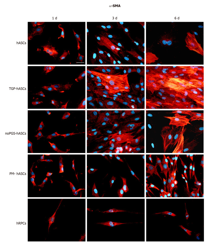
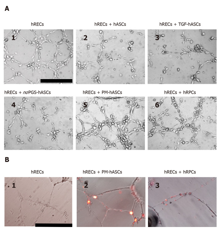
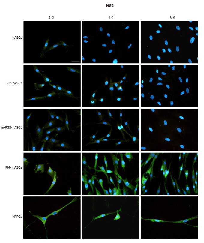
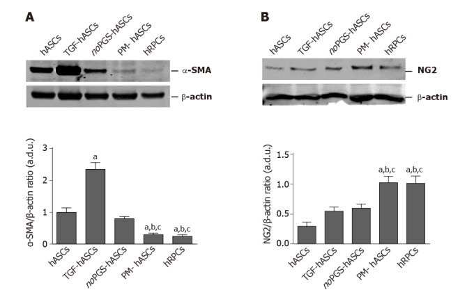
After 3-6 d of culture, α-SMA and NG2 immunocytochemistry showed that the closest pericyte-like phenotype was observed when hASCs were cultured in Pericyte Medium (PM-hASCs). In particular, α-SMA immunoreactivity, already visible at the basal level in pericytes and ASCs, was strongly increased only when transforming growth factor was added to the culture medium. NG2 expression, almost undetectable in most conditions, was substantially increased only in PM-hASCs. Immunocytochemical results were confirmed by western blot analysis. The presence of pericyte growth supplement seems to increase NG2 expression rather than α-SMA, in agreement with its role in maintaining pericytes in the proliferative state. In co-culture experiments, immunoreactivity of vascular endothelial-Cadherin, zonula occludens-1 and Occludin was considerably increased in hRECs when hRPCs or PM-hASCs were also present. Supporting results were found by trans-endothelial electrical resistance measurements, gathered at 3 and 6 d of co-culture. The highest resistance values were obtained when hRECs were co-cultured with hRPCs or PM-hASCs. The pericyte-like phenotype of PM-hASCs was also confirmed in three-dimensional co-cultures in Matrigel, where PM-hASCs and hRPCs similarly localized around the tubular formations made by hRECs.
PM-hASCs seem able to strengthen the intercellular junctions between hRECs, likely reinforcing the BRB; thus, hASC-based therapeutic approaches may be developed to restore the integrity of retinal microcirculation.
脂肪来源的间充质干细胞(ASC)具有长期自我更新和高增殖率的特点。在适当条件下,它们可分化为中胚层、内胚层或外胚层谱系的细胞。周细胞支持内皮细胞,并在微循环水平稳定血管壁方面发挥重要作用。如在糖尿病视网膜病变中发生的周细胞丢失会导致血视网膜屏障(BRB)破坏和炎性细胞浸润。在此背景下,使用类周细胞分化的ASC可能是恢复BRB损伤的一种有价值的治疗策略。
测试使人类ASC(hASC)实现类周细胞分化的策略。
测试了不同的培养条件:在补充有转化生长因子β1的基础培养基中培养的hASC;以及在特定周细胞培养基(PM-hASC)中培养的hASC。在另一个样本中,从PM中省略了周细胞生长补充剂。此外,使用人类视网膜周细胞(hRPC)培养物进行比较。通过免疫细胞化学染色和蛋白质印迹法测试hASC的类周细胞分化,以评估α-平滑肌肌动蛋白(α-SMA)和神经/胶质抗原2(NG2)的表达。在共培养实验中研究了人类视网膜内皮细胞(hREC)与不同组hASC之间的相互作用。在这些情况下,评估hREC中典型连接蛋白如血管内皮钙黏蛋白、闭锁小带-1和闭合蛋白的表达。在BRB模型中,当hREC与各种预处理的hASC组共培养时,测量跨内皮电阻值。将观察到的值与hREC和hRPC的共培养以及单独的hREC培养进行比较。设计了hREC与hRPC或类周细胞hASC在基质胶中的三维共培养,以评估它们的相互定位。
培养3 - 6天后,α-SMA和NG2免疫细胞化学显示,当hASC在周细胞培养基(PM-hASC)中培养时,观察到最接近类周细胞的表型。特别是α-SMA免疫反应性,在周细胞和ASC的基础水平已经可见,仅在向培养基中添加转化生长因子时强烈增加。NG2表达在大多数情况下几乎检测不到,仅在PM-hASC中显著增加。免疫细胞化学结果通过蛋白质印迹分析得到证实。周细胞生长补充剂的存在似乎增加了NG2的表达而非α-SMA,这与其在维持周细胞增殖状态中的作用一致。在共培养实验中,当也存在hRPC或PM-hASC时,hREC中血管内皮钙黏蛋白、闭锁小带-1和闭合蛋白的免疫反应性显著增加。在共培养3天和6天时进行的跨内皮电阻测量得到了支持性结果。当hREC与hRPC或PM-hASC共培养时获得了最高电阻值。PM-hASC的类周细胞表型在基质胶中的三维共培养中也得到了证实,其中PM-hASC和hRPC类似地定位在hREC形成的管状结构周围。
PM-hASC似乎能够加强hREC之间的细胞间连接,可能增强BRB;因此,可开发基于hASC的治疗方法来恢复视网膜微循环的完整性。