• 文献检索
  • 文档翻译
  • 深度研究
  • 学术资讯
  • Suppr Zotero 插件Zotero 插件
  • 邀请有礼
  • 套餐&价格
  • 历史记录
应用&插件
Suppr Zotero 插件Zotero 插件浏览器插件Mac 客户端Windows 客户端微信小程序
定价
高级版会员购买积分包购买API积分包
服务
文献检索文档翻译深度研究API 文档MCP 服务
关于我们
关于 Suppr公司介绍联系我们用户协议隐私条款
关注我们

Suppr 超能文献

核心技术专利:CN118964589B侵权必究
粤ICP备2023148730 号-1Suppr @ 2026

文献检索

告别复杂PubMed语法,用中文像聊天一样搜索,搜遍4000万医学文献。AI智能推荐,让科研检索更轻松。

立即免费搜索

文件翻译

保留排版,准确专业,支持PDF/Word/PPT等文件格式,支持 12+语言互译。

免费翻译文档

深度研究

AI帮你快速写综述,25分钟生成高质量综述,智能提取关键信息,辅助科研写作。

立即免费体验

一项通过磁共振成像和超声检查评估鼻咽癌放疗后患者腮腺和颌下腺变化的纵向研究。

A longitudinal study on parotid and submandibular gland changes assessed by magnetic resonance imaging and ultrasonography in post-radiotherapy nasopharyngeal cancer patients.

作者信息

Wu Vincent W C, Ying Michael Tc, Kwong Dora Lw, Khong Pek-Lan, Wong Gary Kw, Tam Shing-Yau

机构信息

Department of Health Technology and Informatics, Hong Kong Polytechnic University, Hung Hom, Kowloon, Hong Kong.

Department of Clinical Oncology, Li Ka Shing Faculty of Medicine, The University of Hong Kong, Pok Fu Lam, Hong Kong.

出版信息

BJR Open. 2020 Sep 2;2(1):20200003. doi: 10.1259/bjro.20200003. eCollection 2020.

DOI:10.1259/bjro.20200003
PMID:33178971
原文链接:https://pmc.ncbi.nlm.nih.gov/articles/PMC7583169/
Abstract

OBJECTIVES

With regard to the intensity modulated radiotherapy (IMRT) of nasopharyngeal carcinoma (NPC) patients, this longitudinal study evaluated the radiation-induced changes in the parotid and submandibular glands in terms of gland size, echogenicity and haemodynamic parameters.

METHODS

21 NPC patients treated by IMRT underwent MRI and ultrasound scans before radiotherapy, and at 6, 12, 18 and 24 months after treatment. Parotid and submandibular gland volumes were measured from the MRI images, whereas the parotid echogenicity and haemodynamic parameters including the resistive index, pulsatility index, peak systolic velocity and end diastolic velocity were evaluated by ultrasonography. Trend lines were plotted to show the pattern of changes. The correlations of gland doses and the post-RT changes were also studied.

RESULTS

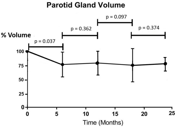
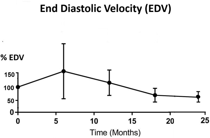
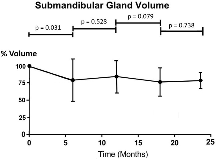
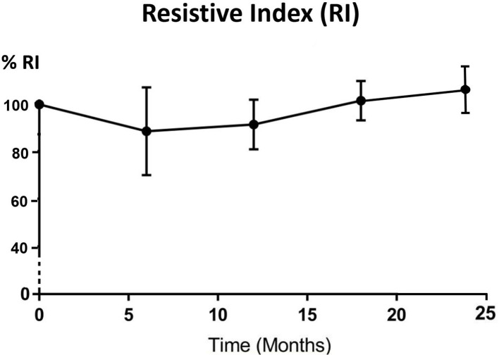
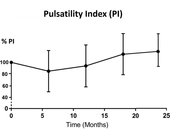
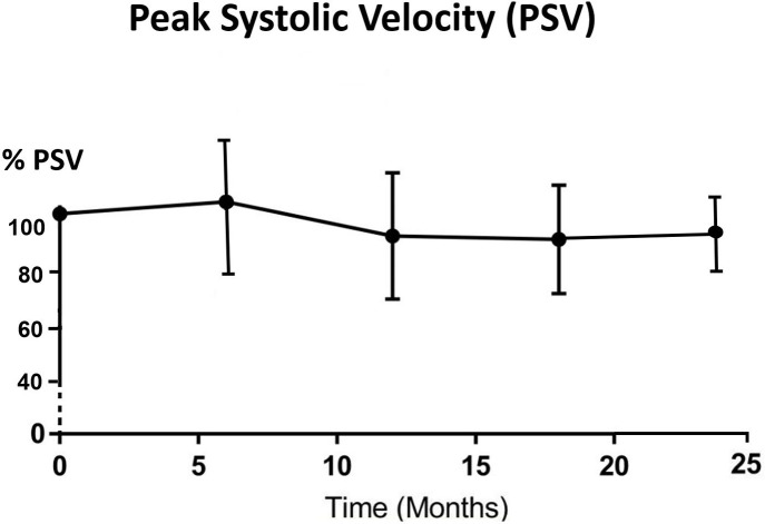
The volume of the parotid and submandibular glands demonstrated a significant drop from pre-RT to 6 months post-RT. The parotid gland changed from hyperechoic before RT to either isoechoic or hypoechoic after treatment. The resistive index and pulsatility index decreased from pre-RT to 6 month post-RT, then started to increase at 12 month time interval. Both peak systolic velocity and end diastolic velocity increased after 6 months post-RT then followed a decreasing trend up to 24 months post-RT. There was mild correlation between post-RT gland dose and gland volume, but not with haemodynamic changes.

CONCLUSIONS

Radiation from IMRT caused shrinkage of parotid and submandibular glands in NPC patients. It also changed the echogenicity and vascular condition of the parotid gland. The most significant changes were observed at 6 months after radiotherapy.

ADVANCES IN KNOWLEDGE

It is the first paper that reports on the longitudinal changes of salivary gland volume, echogenicity and haemodynamic parameters altogether in NPC patients after radiotherapy. The results are useful for the prediction of glandular changes that is associated with xerostomia, which help to provide timely management of the complication when the patients attend follow-up visits.

摘要

目的

针对鼻咽癌(NPC)患者的调强放射治疗(IMRT),本纵向研究从腺体大小、回声性及血流动力学参数方面评估了腮腺和颌下腺的放射性变化。

方法

21例行IMRT治疗的NPC患者在放疗前、治疗后6个月、12个月、18个月及24个月接受了MRI和超声扫描。从MRI图像测量腮腺和颌下腺体积,而通过超声检查评估腮腺回声性及包括阻力指数、搏动指数、收缩期峰值速度和舒张末期速度在内的血流动力学参数。绘制趋势线以显示变化模式。还研究了腺体剂量与放疗后变化的相关性。

结果

腮腺和颌下腺体积从放疗前到放疗后6个月显著下降。腮腺在放疗前为高回声,治疗后变为等回声或低回声。阻力指数和搏动指数从放疗前到放疗后6个月下降,然后在12个月时间间隔开始增加。收缩期峰值速度和舒张末期速度在放疗后6个月后增加,然后在放疗后24个月前呈下降趋势。放疗后腺体剂量与腺体体积之间存在轻度相关性,但与血流动力学变化无关。

结论

IMRT放疗导致NPC患者腮腺和颌下腺萎缩。它还改变了腮腺的回声性和血管状况。放疗后6个月观察到最显著的变化。

知识进展

这是第一篇全面报道NPC患者放疗后唾液腺体积、回声性和血流动力学参数纵向变化的论文。这些结果有助于预测与口干相关的腺体变化,有助于在患者随访时及时处理并发症。

https://cdn.ncbi.nlm.nih.gov/pmc/blobs/3fd1/7583169/08e2e251e8d0/bjro.20200003.g006.jpg
https://cdn.ncbi.nlm.nih.gov/pmc/blobs/3fd1/7583169/f16ddc77bb3a/bjro.20200003.g001.jpg
https://cdn.ncbi.nlm.nih.gov/pmc/blobs/3fd1/7583169/058ba54e110a/bjro.20200003.g002.jpg
https://cdn.ncbi.nlm.nih.gov/pmc/blobs/3fd1/7583169/bad0210d8bc2/bjro.20200003.g003.jpg
https://cdn.ncbi.nlm.nih.gov/pmc/blobs/3fd1/7583169/52e125117112/bjro.20200003.g004.jpg
https://cdn.ncbi.nlm.nih.gov/pmc/blobs/3fd1/7583169/2c37d1f69adb/bjro.20200003.g005.jpg
https://cdn.ncbi.nlm.nih.gov/pmc/blobs/3fd1/7583169/08e2e251e8d0/bjro.20200003.g006.jpg

相似文献

1
A longitudinal study on parotid and submandibular gland changes assessed by magnetic resonance imaging and ultrasonography in post-radiotherapy nasopharyngeal cancer patients.一项通过磁共振成像和超声检查评估鼻咽癌放疗后患者腮腺和颌下腺变化的纵向研究。
BJR Open. 2020 Sep 2;2(1):20200003. doi: 10.1259/bjro.20200003. eCollection 2020.
2
Evaluation of radiation-induced changes to parotid glands following conventional radiotherapy in patients with nasopharygneal carcinoma.评价鼻咽癌患者常规放疗后腮腺的放射性变化。
Br J Radiol. 2011 Sep;84(1005):843-9. doi: 10.1259/bjr/55873561. Epub 2011 Jan 11.
3
Radiation-induced volume changes in parotid and submandibular glands in patients with head and neck cancer receiving postoperative radiotherapy: a longitudinal study.头颈部癌患者术后放疗时腮腺和颌下腺的放射性体积变化:一项纵向研究
Laryngoscope. 2009 Oct;119(10):1966-74. doi: 10.1002/lary.20601.
4
Diffusion-weighted imaging as a follow-up modality for evaluation of major salivary gland function in nasopharyngeal carcinoma patients: a preliminary study.弥散加权成像作为一种随访方式,用于评估鼻咽癌患者大涎腺功能:一项初步研究。
Strahlenther Onkol. 2020 Jun;196(6):530-541. doi: 10.1007/s00066-020-01580-5. Epub 2020 Feb 5.
5
Salivary gland function in nasopharyngeal carcinoma before and late after intensity-modulated radiotherapy evaluated by dynamic diffusion-weighted MR imaging with gustatory stimulation.味觉刺激动态扩散加权 MR 成像评价调强放疗前后鼻咽癌患者唾液腺功能。
BMC Oral Health. 2019 Dec 21;19(1):288. doi: 10.1186/s12903-019-0951-x.
6
Parotid perfusion in nasopharyngeal carcinoma patients in early-to-intermediate stage after low-dose intensity-modulated radiotherapy: evaluated by fat-saturated dynamic contrast-enhanced magnetic resonance imaging.低剂量调强放疗后早中期鼻咽癌患者腮腺灌注:脂肪饱和动态对比增强磁共振成像评估。
Magn Reson Imaging. 2013 Oct;31(8):1278-84. doi: 10.1016/j.mri.2013.03.018. Epub 2013 May 8.
7
Evaluation of Salivary Gland Function Using Diffusion-Weighted Magnetic Resonance Imaging for Follow-Up of Radiation-Induced Xerostomia.使用扩散加权磁共振成像评估涎腺功能对放射性口干症的随访。
Korean J Radiol. 2018 Jul-Aug;19(4):758-766. doi: 10.3348/kjr.2018.19.4.758. Epub 2018 Jun 14.
8
Xerostomia, salivary characteristics and gland volumes following intensity-modulated radiotherapy for nasopharyngeal carcinoma: a two-year follow up.口干症、唾液特性和鼻咽癌调强放疗后腺体体积:两年随访。
Aust Dent J. 2018 Jun;63(2):217-223. doi: 10.1111/adj.12608. Epub 2018 Apr 24.
9
Temporal Evolution of Parotid Volume and Parotid Apparent Diffusion Coefficient in Nasopharyngeal Carcinoma Patients Treated by Intensity-Modulated Radiotherapy Investigated by Magnetic Resonance Imaging: A Pilot Study.磁共振成像研究调强放射治疗鼻咽癌患者腮腺体积及腮腺表观扩散系数的时间演变:一项初步研究
PLoS One. 2015 Aug 31;10(8):e0137073. doi: 10.1371/journal.pone.0137073. eCollection 2015.
10
[The quantitative evaluation of early radiation-induced changes in the salivary glands using MRI].[使用磁共振成像对唾液腺早期辐射诱导变化的定量评估]
Zhonghua Yi Xue Za Zhi. 2017 Feb 21;97(7):492-495. doi: 10.3760/cma.j.issn.0376-2491.2017.07.004.

引用本文的文献

1
Assessment and Prediction of Salivary Gland Function After Head and Neck Radiotherapy: A Systematic Review.头颈部放疗后唾液腺功能的评估与预测:一项系统评价
Cancer Med. 2024 Dec;13(24):e70494. doi: 10.1002/cam4.70494.
2
How Effective Is Radiotherapy in the Ultrasonographic Structural Characteristics of the Submandibular Glands?放射治疗对颌下腺超声结构特征的效果如何?
Eurasian J Med. 2024 Feb 28;56(2):108-113. doi: 10.5152/eurasianjmed.2024.23227.
3
Multi‑parameter quantitative magnetic resonance imaging in the early assessment of radiation‑induced parotid damage in patients with nasopharyngeal carcinoma following intensity‑modulated radiotherapy.

本文引用的文献

1
Spatial Radiation Dose Influence on Xerostomia Recovery and Its Comparison to Acute Incidence in Patients With Head and Neck Cancer.空间辐射剂量对口腔干燥症恢复的影响及其与头颈癌患者急性发病率的比较。
Adv Radiat Oncol. 2019 Aug 31;5(2):221-230. doi: 10.1016/j.adro.2019.08.009. eCollection 2020 Mar-Apr.
2
Predicting acute radiation induced xerostomia in head and neck Cancer using MR and CT Radiomics of parotid and submandibular glands.利用腮腺和颌下腺的 MR 和 CT 影像组学预测头颈部癌症的急性放射性口干症。
Radiat Oncol. 2019 Jul 29;14(1):131. doi: 10.1186/s13014-019-1339-4.
3
Long-term outcomes of interventions for radiation-induced xerostomia: A review.
多参数定量磁共振成像在鼻咽癌调强放疗后放射性腮腺损伤早期评估中的应用
Oncol Lett. 2024 Feb 28;27(4):180. doi: 10.3892/ol.2024.14313. eCollection 2024 Apr.
4
Early Experience of Online Adaptive Radiation Therapy for Definitive Radiation of Patients With Head and Neck Cancer.头颈部癌患者确定性放疗的在线自适应放射治疗早期经验
Adv Radiat Oncol. 2023 Apr 26;8(5):101256. doi: 10.1016/j.adro.2023.101256. eCollection 2023 Sep-Oct.
5
Structural network alterations in patients with nasopharyngeal carcinoma after radiotherapy: A 1-year longitudinal study.鼻咽癌患者放疗后结构网络改变:一项为期1年的纵向研究。
Front Neurosci. 2022 Nov 17;16:1059320. doi: 10.3389/fnins.2022.1059320. eCollection 2022.
6
Research Value of Intensity Modulated Radiation Therapy in Alleviating Parotid Gland Function Injury in Patients with Stage N0 Nasopharyngeal Carcinoma from Physical and Dosimetric Aspects.从物理和剂量学方面评估调强放疗减轻 N0 期鼻咽癌患者腮腺功能损伤的研究价值。
Comput Math Methods Med. 2022 Jul 11;2022:4651364. doi: 10.1155/2022/4651364. eCollection 2022.
7
Current Omics Trends in Personalised Head and Neck Cancer Chemoradiotherapy.个性化头颈癌放化疗中的当前组学趋势
J Pers Med. 2021 Oct 26;11(11):1094. doi: 10.3390/jpm11111094.
放射性口干症干预措施的长期效果:一项综述。
World J Clin Oncol. 2019 Jan 10;10(1):1-13. doi: 10.5306/wjco.v10.i1.1.
4
Parotid gland fat related Magnetic Resonance image biomarkers improve prediction of late radiation-induced xerostomia.腮腺脂肪相关磁共振成像生物标志物可改善晚期放射性口干症的预测。
Radiother Oncol. 2018 Sep;128(3):459-466. doi: 10.1016/j.radonc.2018.06.012. Epub 2018 Jun 26.
5
Parotid gland radiation dose-xerostomia relationships based on actual delivered dose for nasopharyngeal carcinoma.基于鼻咽癌实际照射剂量的腮腺辐射剂量与口干症的关系
J Appl Clin Med Phys. 2018 May;19(3):251-260. doi: 10.1002/acm2.12327. Epub 2018 Apr 17.
6
Xerostomia, salivary characteristics and gland volumes following intensity-modulated radiotherapy for nasopharyngeal carcinoma: a two-year follow up.口干症、唾液特性和鼻咽癌调强放疗后腺体体积:两年随访。
Aust Dent J. 2018 Jun;63(2):217-223. doi: 10.1111/adj.12608. Epub 2018 Apr 24.
7
Design and Selection of Machine Learning Methods Using Radiomics and Dosiomics for Normal Tissue Complication Probability Modeling of Xerostomia.使用放射组学和剂量组学进行机器学习方法的设计与选择,以建立口干症正常组织并发症概率模型
Front Oncol. 2018 Mar 5;8:35. doi: 10.3389/fonc.2018.00035. eCollection 2018.
8
F-FDG PET image biomarkers improve prediction of late radiation-induced xerostomia.18F-FDG PET 图像生物标志物可改善晚期放射性口干的预测。
Radiother Oncol. 2018 Jan;126(1):89-95. doi: 10.1016/j.radonc.2017.08.024. Epub 2017 Sep 23.
9
Clinical study of the time of repeated computed tomography and replanning for patients with nasopharyngeal carcinoma.鼻咽癌患者重复计算机断层扫描时间及重新计划的临床研究
Oncotarget. 2017 Apr 18;8(16):27529-27540. doi: 10.18632/oncotarget.16770.
10
A longitudinal evaluation of early anatomical changes of parotid gland in intensity modulated radiotherapy of nasopharyngeal carcinoma patients with parapharyngeal space involvement.鼻咽癌累及咽旁间隙患者调强放疗中腮腺早期解剖学变化的纵向评估
J Med Radiat Sci. 2017 Sep;64(3):188-194. doi: 10.1002/jmrs.209. Epub 2017 Mar 4.