Department of Physiology, Hubei Key Laboratory of Embryonic Stem Cell Research, School of Basic Medicine Science, Hubei University of Medicine, 442000, Hubei, China.
Institute of Biomedicine, Hubei University of Medicine, 442000, Hubei, China.
Cell Death Dis. 2020 Nov 12;11(11):971. doi: 10.1038/s41419-020-03142-0.
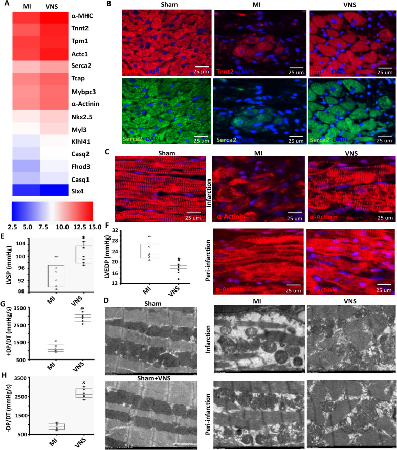
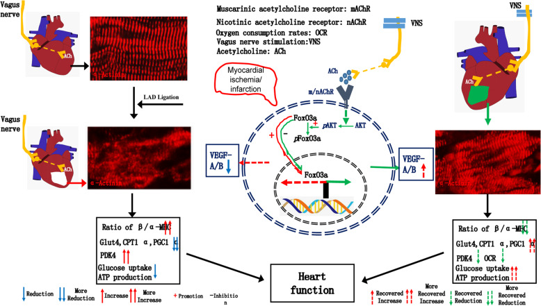
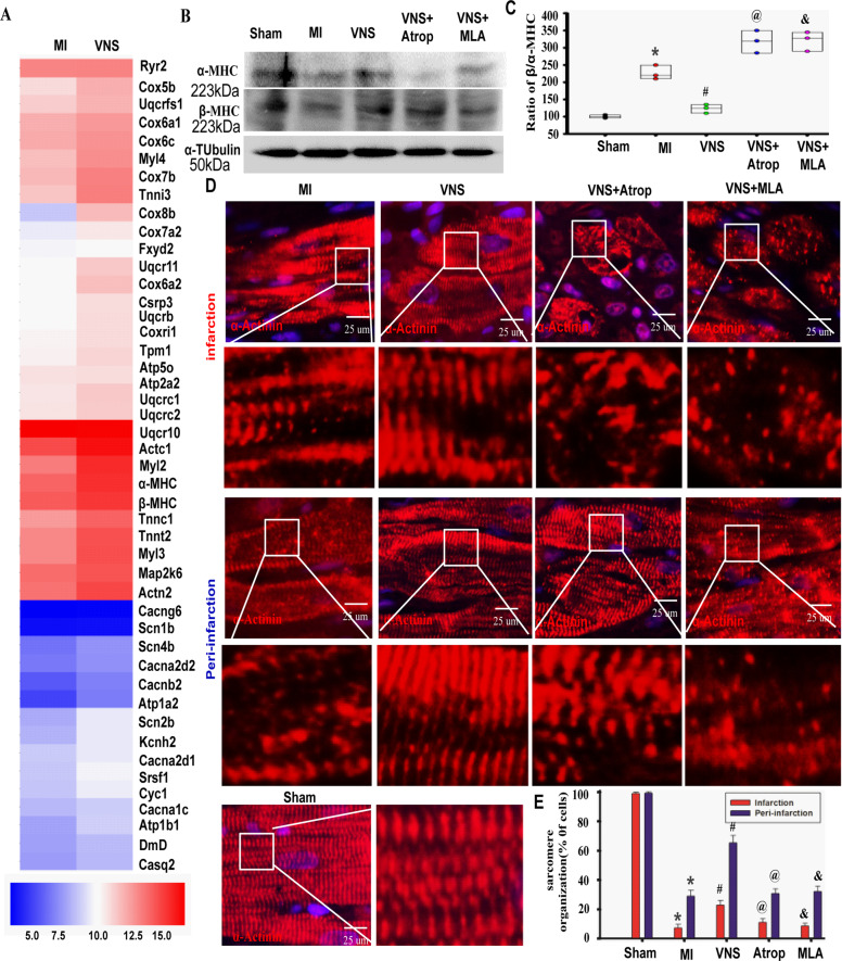
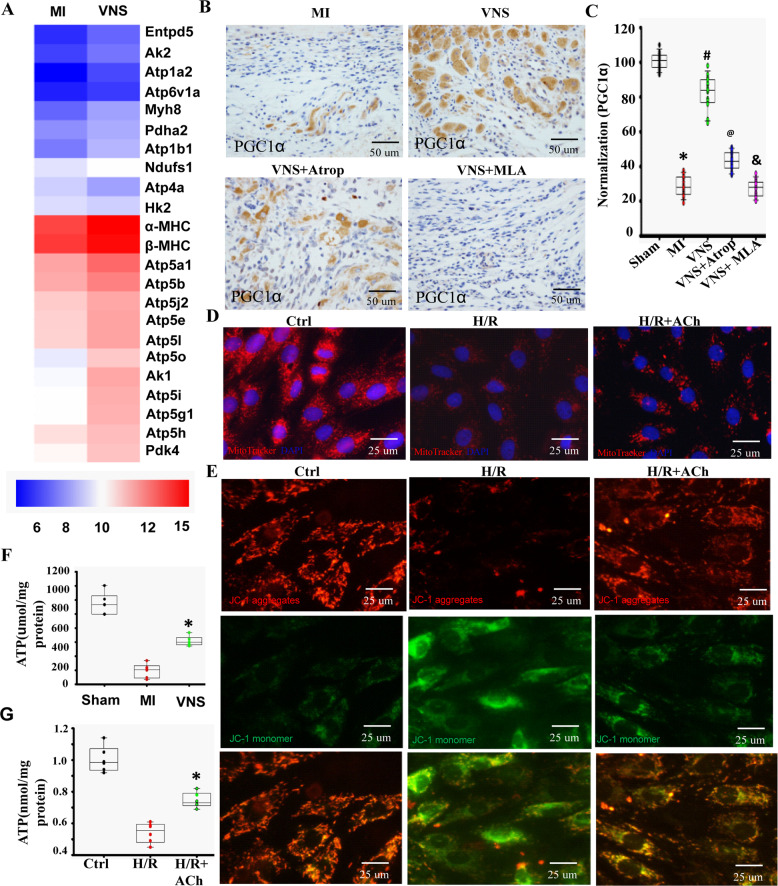
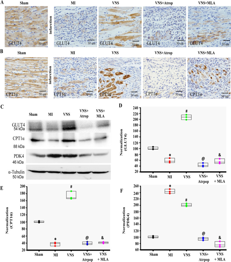
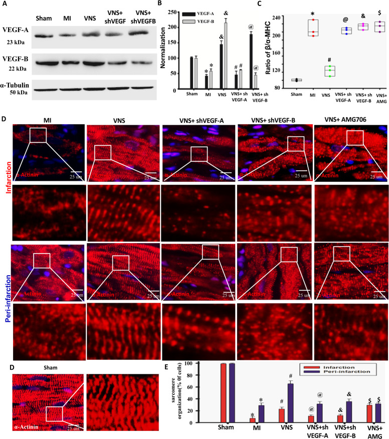
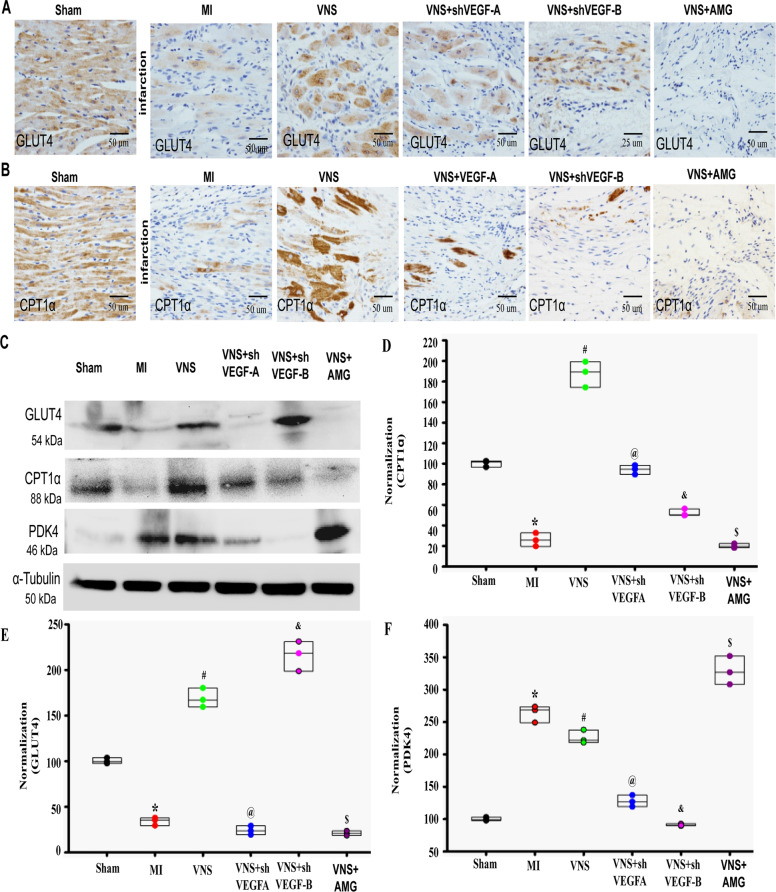
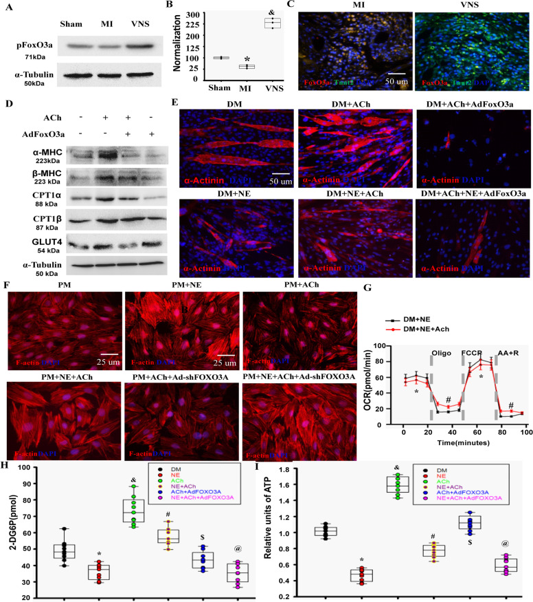
Vagus nerve stimulation (VNS) restores autonomic balance, suppresses inflammation action and minimizes cardiomyocyte injury. However, little knowledge is known about the VNS' role in cardiomyocyte phenotype, sarcomere organization, and energy metabolism of infarcted hearts. VNS in vivo and acetylcholine (ACh) in vitro optimized the levels of α/β-MHC and α-Actinin positive sarcomere organization in cardiomyocytes while reducing F-actin assembly of cardiomyocytes. Consistently, ACh improved glucose uptake while decreasing lipid deposition in myocytes, correlating both with the increase of Glut4 and CPT1α and the decrease of PDK4 in infarcted hearts in vivo and myocytes in vitro, attributing to improvement in both glycolysis by VEGF-A and lipid uptake by VEGF-B in response to Ach. This led to increased ATP levels accompanied by the repaired mitochondrial function and the decreased oxygen consumption. Functionally, VNS improved the left ventricular performance. In contrast, ACh-m/nAChR inhibitor or knockdown of VEGF-A/B by shRNA powerfully abrogated these effects mediated by VNS. On mechanism, ACh decreased the levels of nuclear translocation of FoxO3A in myocytes due to phosphorylation of FoxO3A by activating AKT. FoxO3A overexpression or knockdown could reverse the specific effects of ACh on the expression of VEGF-A/B, α/β-MHC, Glut4, and CPT1α, sarcomere organization, glucose uptake and ATP production. Taken together, VNS optimized cardiomyocytes sarcomere organization and energy metabolism to improve heart function of the infarcted heart during the process of delaying and/or blocking the switch from compensated hypertrophy to decompensated heart failure, which were associated with activation of both P13K/AKT-FoxO3A-VEGF-A/B signaling cascade.
迷走神经刺激 (VNS) 可恢复自主平衡,抑制炎症作用,并最大限度地减少心肌细胞损伤。然而,对于 VNS 在梗塞心脏中心肌细胞表型、肌节组织和能量代谢中的作用知之甚少。体内的 VNS 和体外的乙酰胆碱 (ACh) 优化了心肌细胞中 α/β-MHC 和 α-肌球蛋白重链阳性肌节组织的水平,同时减少了心肌细胞中 F-肌动蛋白的组装。一致地,ACh 增加了葡萄糖摄取,同时减少了肌细胞中的脂质沉积,这与体内梗塞心脏和体外心肌细胞中 Glut4 和 CPT1α 的增加以及 PDK4 的减少相关,这归因于 VEGF-A 对糖酵解和 VEGF-B 对脂质摄取的改善。这导致 ATP 水平升高,同时线粒体功能得到修复,耗氧量降低。功能上,VNS 改善了左心室功能。相比之下,ACh-m/nAChR 抑制剂或通过 shRNA 敲低 VEGF-A/B 有力地削弱了 VNS 介导的这些作用。在机制上,由于激活 AKT 使 FoxO3A 磷酸化,ACh 降低了肌细胞中核转位 FoxO3A 的水平。FoxO3A 过表达或敲低可以逆转 ACh 对 VEGF-A/B、α/β-MHC、Glut4 和 CPT1α 的表达、肌节组织、葡萄糖摄取和 ATP 产生的特定作用。总之,VNS 优化了心肌细胞肌节组织和能量代谢,以改善梗塞心脏的心脏功能,在从代偿性肥大到失代偿性心力衰竭的转变过程中延迟和/或阻断这种转变,这与 P13K/AKT-FoxO3A-VEGF-A/B 信号级联的激活有关。