• 文献检索
  • 文档翻译
  • 深度研究
  • 学术资讯
  • Suppr Zotero 插件Zotero 插件
  • 邀请有礼
  • 套餐&价格
  • 历史记录
应用&插件
Suppr Zotero 插件Zotero 插件浏览器插件Mac 客户端Windows 客户端微信小程序
定价
高级版会员购买积分包购买API积分包
服务
文献检索文档翻译深度研究API 文档MCP 服务
关于我们
关于 Suppr公司介绍联系我们用户协议隐私条款
关注我们

Suppr 超能文献

核心技术专利:CN118964589B侵权必究
粤ICP备2023148730 号-1Suppr @ 2026

文献检索

告别复杂PubMed语法,用中文像聊天一样搜索,搜遍4000万医学文献。AI智能推荐,让科研检索更轻松。

立即免费搜索

文件翻译

保留排版,准确专业,支持PDF/Word/PPT等文件格式,支持 12+语言互译。

免费翻译文档

深度研究

AI帮你快速写综述,25分钟生成高质量综述,智能提取关键信息,辅助科研写作。

立即免费体验

测量与年龄相关性黄斑变性相关的基底膜和布鲁赫膜的贡献。

Measuring the Contributions of Basal Laminar Deposit and Bruch's Membrane in Age-Related Macular Degeneration.

机构信息

Department of Ophthalmology and Visual Sciences, University of Alabama at Birmingham School of Medicine, Birmingham, Alabama, United States.

The First Affiliated Hospital of Chongqing Medical University, Chongqing Key Laboratory of Ophthalmology, and Chongqing Eye Institute, Chongqing, China.

出版信息

Invest Ophthalmol Vis Sci. 2020 Nov 2;61(13):19. doi: 10.1167/iovs.61.13.19.

DOI:10.1167/iovs.61.13.19
PMID:33186466
原文链接:https://pmc.ncbi.nlm.nih.gov/articles/PMC7671869/
Abstract

PURPOSE

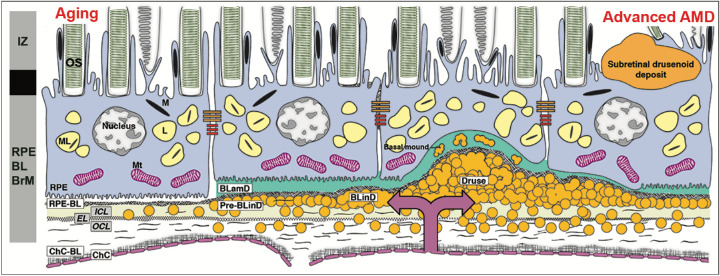
Basal laminar deposit (BLamD) is a consistent finding in age-related macular degeneration (AMD). We quantified BLamD thickness, appearance, and topography in eyes of aged donors with and without AMD and evaluated its relationship to other components of the retinal pigment epithelium-basal lamina/Bruch's membrane (RPE-BL-BrM) complex.

METHODS

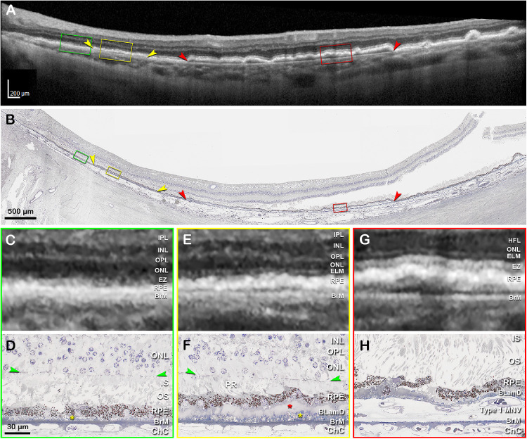
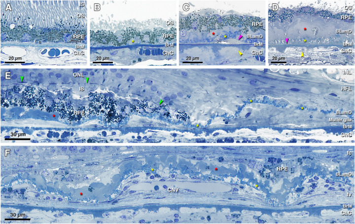
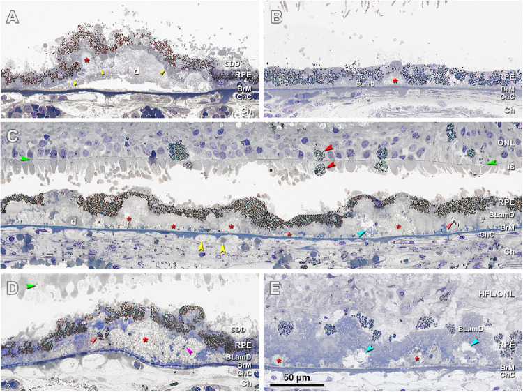
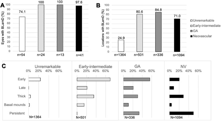
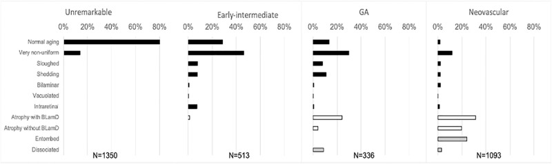
Donor eyes (n = 132) were classified as normal (n = 54), early to intermediate AMD (n = 24), geographic atrophy (GA; n = 13), and neovascular AMD (NV; n = 41). In high-resolution histology, we assessed RPE, BLamD, and BrM thicknesses and phenotypes at 3309 predefined locations in the central (foveal and perifovea) and superior (perifoveal) sections. Pre-mortem optical coherence tomography (OCT) imaging of a 90-year-old woman was compared to postmortem histopathology.

RESULTS

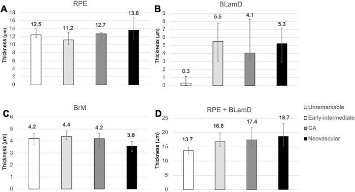
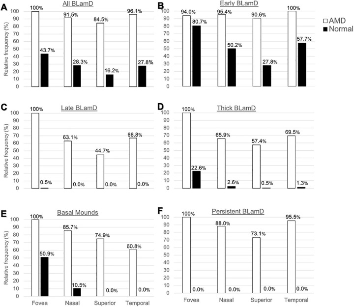
In non-atrophic areas of AMD eyes, the RPE-BLamD is thick (normal = 13.7 µm, early-intermediate = 16.8 µm, GA = 17.4 µm, NV = 18.7 µm), because the BLamD is thick (normal = 0.3 µm, early-intermediate = 5.5 µm, GA = 4.1 µm, NV = 5.3 µm). RPE layer thickness is similar across these stages. Disease-associated variants of BLamD (thick, late, basal mounds) cluster subfoveally. A thick BLamD is visible on OCT as a hyporeflective split in the RPE-BL-BrM complex. BrM is thin (3.5 µm) in NV (normal = 4.2 µm, early to intermediate = 4.4 µm, and GA = 4.2 µm).

CONCLUSIONS

The RPE-BL-BrM complex is thick in AMD, driven by the accumulation and expansion of BLamD rather than expansion of either three-layer BrM, RPE-BL, or RPE. BLamD is clinically appreciable by OCT in some patients as a non-neovascular "split RPE-BL-BrM complex" or "double-layer sign." BLamD may contribute toward the formation and progression of high-risk drusen yet also exhibit protective properties.

摘要

目的

基底膜层沉积物(BLamD)是年龄相关性黄斑变性(AMD)的一种常见表现。我们定量分析了 AMD 眼和非 AMD 眼的 BLamD 厚度、外观和分布,并评估了其与视网膜色素上皮-基底膜/布鲁赫膜(RPE-BL-BrM)复合体其他成分的关系。

方法

对 132 只供体眼(n=132)进行分类:正常(n=54)、早期至中期 AMD(n=24)、地图状萎缩(GA;n=13)和新生血管性 AMD(NV;n=41)。在高分辨率组织学中,我们在中央(中心和旁中心)和上(旁中心)区域的 3309 个预设位置评估了 RPE、BLamD 和 BrM 厚度和表型。对一名 90 岁女性的生前光学相干断层扫描(OCT)成像与死后组织病理学进行了比较。

结果

在 AMD 眼的非萎缩区域,RPE-BLamD 较厚(正常=13.7μm,早期-中期=16.8μm,GA=17.4μm,NV=18.7μm),因为 BLamD 较厚(正常=0.3μm,早期-中期=5.5μm,GA=4.1μm,NV=5.3μm)。这些阶段的 RPE 层厚度相似。与疾病相关的 BLamD 变体(厚、晚期、基底丘)聚集在中心下。OCT 上可见 BLamD 增厚,表现为 RPE-BL-BrM 复合体的低反射性分离。BrM 在 NV 中较薄(3.5μm)(正常=4.2μm,早期-中期=4.4μm,GA=4.2μm)。

结论

AMD 中 RPE-BL-BrM 复合体较厚,由 BLamD 的积累和扩张引起,而不是三层 BrM、RPE-BL 或 RPE 的扩张。在一些患者中,BLamD 可通过 OCT 临床识别,表现为非新生血管性“RPE-BL-BrM 复合体分离”或“双层征”。BLamD 可能有助于高危 drusen 的形成和进展,但也表现出保护特性。

https://cdn.ncbi.nlm.nih.gov/pmc/blobs/7de8/7671869/cb35204b39aa/iovs-61-13-19-f008.jpg
https://cdn.ncbi.nlm.nih.gov/pmc/blobs/7de8/7671869/1dc5ef6fef7e/iovs-61-13-19-f001.jpg
https://cdn.ncbi.nlm.nih.gov/pmc/blobs/7de8/7671869/25fddfca61fd/iovs-61-13-19-f002.jpg
https://cdn.ncbi.nlm.nih.gov/pmc/blobs/7de8/7671869/281e3d866558/iovs-61-13-19-f003.jpg
https://cdn.ncbi.nlm.nih.gov/pmc/blobs/7de8/7671869/cfc91fa02c73/iovs-61-13-19-f004.jpg
https://cdn.ncbi.nlm.nih.gov/pmc/blobs/7de8/7671869/03263e295d50/iovs-61-13-19-f005.jpg
https://cdn.ncbi.nlm.nih.gov/pmc/blobs/7de8/7671869/05e5ffccd644/iovs-61-13-19-f006.jpg
https://cdn.ncbi.nlm.nih.gov/pmc/blobs/7de8/7671869/ff5091d5ccd8/iovs-61-13-19-f007.jpg
https://cdn.ncbi.nlm.nih.gov/pmc/blobs/7de8/7671869/cb35204b39aa/iovs-61-13-19-f008.jpg

相似文献

1
Measuring the Contributions of Basal Laminar Deposit and Bruch's Membrane in Age-Related Macular Degeneration.测量与年龄相关性黄斑变性相关的基底膜和布鲁赫膜的贡献。
Invest Ophthalmol Vis Sci. 2020 Nov 2;61(13):19. doi: 10.1167/iovs.61.13.19.
2
The Border of Macular Atrophy in Age-Related Macular Degeneration: A Clinicopathologic Correlation.年龄相关性黄斑变性中黄斑萎缩边界:临床病理相关性。
Am J Ophthalmol. 2018 Sep;193:166-177. doi: 10.1016/j.ajo.2018.06.020. Epub 2018 Jul 5.
3
Subducted and melanotic cells in advanced age-related macular degeneration are derived from retinal pigment epithelium.年龄相关性黄斑变性晚期的内陷和黑素化细胞源自视网膜色素上皮。
Invest Ophthalmol Vis Sci. 2015 May;56(5):3269-78. doi: 10.1167/iovs.15-16432.
4
Topographic Measurement of the Subretinal Pigment Epithelium Space in Normal Aging and Age-Related Macular Degeneration Using High-Resolution OCT.使用高分辨率 OCT 对正常衰老和年龄相关性黄斑变性的视网膜色素上皮下空间进行地形测量。
Invest Ophthalmol Vis Sci. 2024 Aug 1;65(10):18. doi: 10.1167/iovs.65.10.18.
5
VISUALIZING RETINAL PIGMENT EPITHELIUM PHENOTYPES IN THE TRANSITION TO ATROPHY IN NEOVASCULAR AGE-RELATED MACULAR DEGENERATION.在新生血管性年龄相关性黄斑变性向萎缩转变过程中可视化视网膜色素上皮细胞表型
Retina. 2016 Dec;36 Suppl 1(Suppl 1):S26-S39. doi: 10.1097/IAE.0000000000001330.
6
VISUALIZING RETINAL PIGMENT EPITHELIUM PHENOTYPES IN THE TRANSITION TO GEOGRAPHIC ATROPHY IN AGE-RELATED MACULAR DEGENERATION.在年龄相关性黄斑变性向地图样萎缩转变过程中可视化视网膜色素上皮表型
Retina. 2016 Dec;36 Suppl 1(Suppl 1):S12-S25. doi: 10.1097/IAE.0000000000001276.
7
Ultrahigh Resolution OCT Markers of Normal Aging and Early Age-related Macular Degeneration.正常衰老和早期年龄相关性黄斑变性的超高分辨率光学相干断层扫描标志物
Ophthalmol Sci. 2023 Feb 2;3(3):100277. doi: 10.1016/j.xops.2023.100277. eCollection 2023 Sep.
8
Histologic and morphometric analysis of the choroid, Bruch's membrane, and retinal pigment epithelium in postmortem eyes with age-related macular degeneration and histologic examination of surgically excised choroidal neovascular membranes.对患有年龄相关性黄斑变性的死后眼球的脉络膜、布鲁赫膜和视网膜色素上皮进行组织学和形态计量学分析,并对手术切除的脉络膜新生血管膜进行组织学检查。
Surv Ophthalmol. 1999 Oct;44 Suppl 1:S10-32. doi: 10.1016/s0039-6257(99)00086-7.
9
Relationship of Basal laminar deposit and membranous debris to the clinical presentation of early age-related macular degeneration.基底膜沉积物和膜性碎片与早发性年龄相关性黄斑变性临床表现的关系。
Invest Ophthalmol Vis Sci. 2007 Mar;48(3):968-77. doi: 10.1167/iovs.06-0443.
10
The Project MACULA Retinal Pigment Epithelium Grading System for Histology and Optical Coherence Tomography in Age-Related Macular Degeneration.年龄相关性黄斑变性组织学和光学相干断层扫描的黄斑视网膜色素上皮分级系统项目
Invest Ophthalmol Vis Sci. 2015 May;56(5):3253-68. doi: 10.1167/iovs.15-16431.

引用本文的文献

1
Progression Patterns in Foveal-Sparing Geographic Atrophy With Double-Layer Sign Due to Neovascularization or Basal Laminar Deposits.因新生血管或基底膜沉积物导致的具有双层征的黄斑中心凹保留型地图样萎缩的进展模式。
Invest Ophthalmol Vis Sci. 2025 Aug 1;66(11):16. doi: 10.1167/iovs.66.11.16.
2
En Face OCT and the Phenotype Characterization of Drusen.正面光学相干断层扫描与玻璃膜疣的表型特征
Invest Ophthalmol Vis Sci. 2025 Jul 1;66(9):52. doi: 10.1167/iovs.66.9.52.
3
Genetic Associations of Rod- and Cone-Mediated Vision in Aging and Age-Related Macular Degeneration.

本文引用的文献

1
Visualizing human photoreceptor and retinal pigment epithelium cell mosaics in a single volume scan over an extended field of view with adaptive optics optical coherence tomography.利用自适应光学光学相干断层扫描技术在扩展视野的单次容积扫描中可视化人类光感受器和视网膜色素上皮细胞镶嵌图。
Biomed Opt Express. 2020 Jul 22;11(8):4520-4535. doi: 10.1364/BOE.393906. eCollection 2020 Aug 1.
2
High-Speed, Ultrahigh-Resolution Spectral-Domain OCT with Extended Imaging Range Using Reference Arm Length Matching.高速、超高分辨率谱域 OCT 采用参考臂长匹配技术扩展成像范围。
Transl Vis Sci Technol. 2020 Jun 8;9(7):12. doi: 10.1167/tvst.9.7.12. eCollection 2020 Jun.
3
衰老及年龄相关性黄斑变性中视杆和视锥介导视觉的遗传关联
Invest Ophthalmol Vis Sci. 2025 Jul 1;66(9):50. doi: 10.1167/iovs.66.9.50.
4
In Vivo Characterization of Bruch's Membrane Breaks in Neovascular Age-Related Macular Degeneration Using High-Resolution Optical Coherence Tomography.使用高分辨率光学相干断层扫描对新生血管性年龄相关性黄斑变性中 Bruch 膜破裂的体内特征分析
Invest Ophthalmol Vis Sci. 2025 Jul 1;66(9):19. doi: 10.1167/iovs.66.9.19.
5
Histological Assessment of Foveal Mechanical Instability and Potential Implications for Macular Diseases.黄斑中心凹机械性不稳定的组织学评估及其对黄斑疾病的潜在影响
Invest Ophthalmol Vis Sci. 2025 Jun 2;66(6):82. doi: 10.1167/iovs.66.6.82.
6
Targeting Senescence, Oxidative Stress, and Inflammation: Quercetin-Based Strategies for Ocular Diseases in Older Adults.针对衰老、氧化应激和炎症:基于槲皮素的老年人眼部疾病治疗策略。
Clin Interv Aging. 2025 Jun 7;20:791-813. doi: 10.2147/CIA.S516946. eCollection 2025.
7
Quantifications of Outer Retinal Bands in Geographic Atrophy by Comparing Superior Axial Resolution and Conventional OCT.通过比较上轴分辨率和传统光学相干断层扫描对地图样萎缩中视网膜外层带进行定量分析。
Invest Ophthalmol Vis Sci. 2025 Apr 1;66(4):65. doi: 10.1167/iovs.66.4.65.
8
Topographic Analysis of Two-Wavelength Autofluorescence Supports Higher Macular Xanthophyll Pigment in AMD Than Aging: ALSTAR2 Baseline.两波长自体荧光的地形图分析表明,与衰老相比,年龄相关性黄斑变性患者黄斑区叶黄素色素含量更高:ALSTAR2基线研究。
Invest Ophthalmol Vis Sci. 2025 Mar 3;66(3):61. doi: 10.1167/iovs.66.3.61.
9
Dissecting the biological complexity of age-related macular degeneration: Is it one disease, multiple separate diseases, or a spectrum?剖析年龄相关性黄斑变性的生物学复杂性:它是一种疾病、多种独立疾病,还是一个疾病谱?
Exp Eye Res. 2025 May;254:110304. doi: 10.1016/j.exer.2025.110304. Epub 2025 Feb 19.
10
RPE Basal Lamina Biology and Pathophysiology Related to Age-Related Macular Degeneration.与年龄相关性黄斑变性相关的视网膜色素上皮基底膜生物学与病理生理学
Adv Exp Med Biol. 2025;1468:15-19. doi: 10.1007/978-3-031-76550-6_3.
Atlas of Human Retinal Pigment Epithelium Organelles Significant for Clinical Imaging.
人类视网膜色素上皮细胞器图谱:对临床成像有重要意义。
Invest Ophthalmol Vis Sci. 2020 Jul 1;61(8):13. doi: 10.1167/iovs.61.8.13.
4
Characterization of Drusen and Hyperreflective Foci as Biomarkers for Disease Progression in Age-Related Macular Degeneration Using Artificial Intelligence in Optical Coherence Tomography.利用人工智能在光学相干断层扫描中对年龄相关性黄斑变性的特征进行分析,以评估其作为疾病进展的生物标志物。
JAMA Ophthalmol. 2020 Jul 1;138(7):740-747. doi: 10.1001/jamaophthalmol.2020.1376.
5
Nonexudative Macular Neovascularization Supporting Outer Retina in Age-Related Macular Degeneration: A Clinicopathologic Correlation.年龄相关性黄斑变性中支持外视网膜的非渗出性黄斑新生血管:临床病理相关性。
Ophthalmology. 2020 Jul;127(7):931-947. doi: 10.1016/j.ophtha.2020.01.040. Epub 2020 Jan 29.
6
Long-term Evolution and Remodeling of Soft Drusen in Rhesus Macaques.软玻璃体斑在恒河猴中的长期演变和重塑。
Invest Ophthalmol Vis Sci. 2020 Feb 7;61(2):32. doi: 10.1167/iovs.61.2.32.
7
Consensus Nomenclature for Reporting Neovascular Age-Related Macular Degeneration Data: Consensus on Neovascular Age-Related Macular Degeneration Nomenclature Study Group.《关于新生血管性年龄相关性黄斑变性数据报告的共识命名法:新生血管性年龄相关性黄斑变性命名法研究组的共识》。
Ophthalmology. 2020 May;127(5):616-636. doi: 10.1016/j.ophtha.2019.11.004. Epub 2019 Nov 14.
8
SUBRETINAL DRUSENOID DEPOSIT IN AGE-RELATED MACULAR DEGENERATION: Histologic Insights Into Initiation, Progression to Atrophy, and Imaging.年龄相关性黄斑变性的视网膜下硬性脂褐质沉积:发病机制、向萎缩进展及影像学研究的组织学观察。
Retina. 2020 Apr;40(4):618-631. doi: 10.1097/IAE.0000000000002657.
9
Late-onset retinal degeneration pathology due to mutations in CTRP5 is mediated through HTRA1.由CTRP5突变引起的迟发性视网膜变性病理是通过HTRA1介导的。
Aging Cell. 2019 Dec;18(6):e13011. doi: 10.1111/acel.13011. Epub 2019 Aug 5.
10
CLINICOPATHOLOGIC CORRELATION OF GEOGRAPHIC ATROPHY SECONDARY TO AGE-RELATED MACULAR DEGENERATION.年龄相关性黄斑变性所致地图状萎缩的临床病理相关性。
Retina. 2019 Apr;39(4):802-816. doi: 10.1097/IAE.0000000000002461.