Fragiotta Serena, Parravano Mariacristina, Sacconi Riccardo, Polito Maria Sole, Cioffi Benedetta, Rissotto Federico, Beretta Federico, Costanzo Eliana, Romano Enrico, Capuano Vittorio, Souied Eric H, Querques Giuseppe
Ophthalmology Unit, "Sapienza" University of Rome, NESMOS Department, St. Andrea Hospital, Rome, Italy.
IRCCS-Fondazione Bietti, Rome, Italy.
Invest Ophthalmol Vis Sci. 2025 Aug 1;66(11):16. doi: 10.1167/iovs.66.11.16.
The purpose of this study was to analyze the prognostic significance of double-layer sign (DLS) in eyes with foveal-sparing geographic atrophy (GA) secondary to age-related macular degeneration.
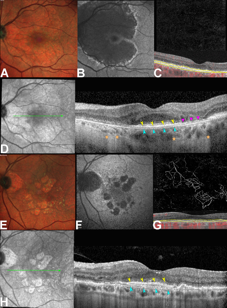
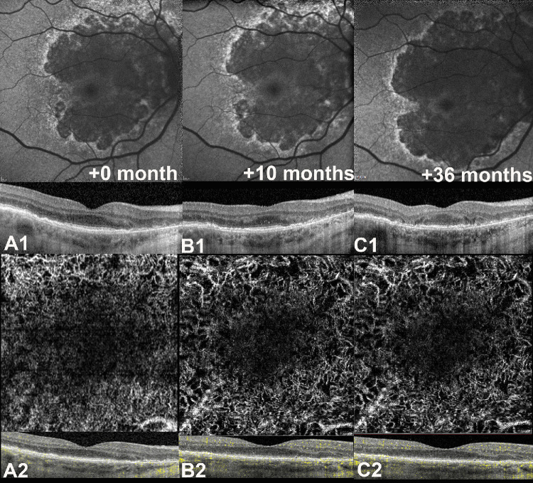
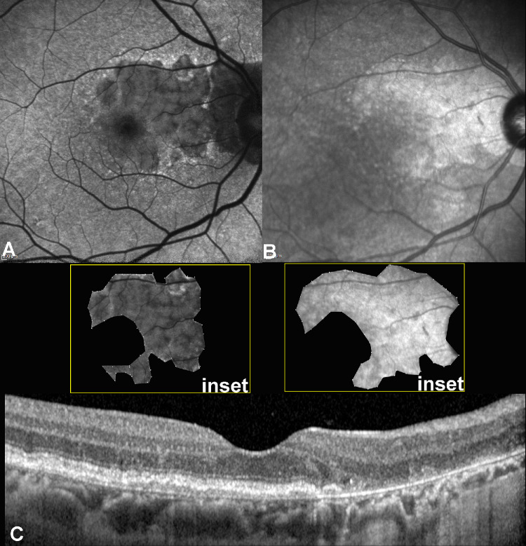
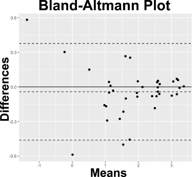
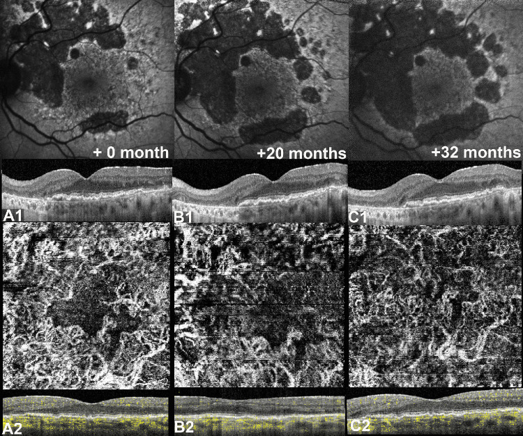
This retrospective, observational cohort study analyzed 46 eyes (46 patients) with foveal sparing GA and associated DLS, using fundus autofluorescence (FAF), near-infrared reflectance (NIR), optical coherence tomography (OCT), and OCT angiography (OCTA). DLS was defined based on OCTA findings as either thick basal laminar deposits (BLamD) or non-exudative macular neovascularization (NE-MNV). The area of GA and foveal sparing were estimated on both FAF and NIR at different time points. Centrifugal and centripetal GA growth rates referring to the lesion expansion away from and toward the fovea, respectively, were evaluated using a mathematical formula.
Of the 46 eyes enrolled, 25 had thick BLamD, whereas 21 had type 1 NE-MNV. The NE-MNV eyes showed significantly thicker DLS than those with BLamD (90.4 ± 39.8 µm vs. 57.0 ± 27 µm, 95% confidence interval [CI] = 0.34 to 0.78, P < 0.001). GA areas were smaller on FAF than NIR (95% CI = -0.89 to -0.03, P = 0.03) in the BLamD group, whereas no difference was observed in the NE-MNV group (95% CI = -0.37 to 0.64, P = 0.60). Despite similar GA areas, the NE-MNV eyes exhibited larger foveal sparing (95% CI = 0.02 to 1.21, P = 0.04). The foveal sparing area remained stable (F(1.2, 11 = 4.15, P = 0.06, ω2 = 0.02) in the NE-MNV group, whereas a significant reduction was observed in the BLamD subgroup (F(1.39, 20.9) = 7.5, P < 0.001, ω2 = 0.09).
OCTA has provided valuable insights into the pathogenic interpretation of the DLS signature. Our findings confirm that a neovascular DLS protects the retinal pigment epithelium and outer retina, contributing to prolonged foveal preservation.
本研究旨在分析双层征(DLS)在年龄相关性黄斑变性继发的保留中心凹的地图样萎缩(GA)眼中的预后意义。
这项回顾性观察性队列研究分析了46只患有保留中心凹GA并伴有DLS的眼睛(46例患者),使用了眼底自发荧光(FAF)、近红外反射(NIR)、光学相干断层扫描(OCT)和OCT血管造影(OCTA)。根据OCTA结果,DLS被定义为厚的基底膜沉积物(BLamD)或非渗出性黄斑新生血管(NE-MNV)。在不同时间点,通过FAF和NIR评估GA和中心凹保留的面积。使用数学公式分别评估离心性和向心性GA增长率,分别指病变远离和朝向中心凹的扩展。
在纳入的46只眼中,25只患有厚的BLamD,而21只患有1型NE-MNV。NE-MNV眼的DLS明显比BLamD眼厚(90.4±39.8μm对57.0±27μm,95%置信区间[CI]=0.34至0.78,P<0.001)。在BLamD组中,FAF上的GA面积比NIR上小(95%CI=-0.89至-0.03,P=0.03),而在NE-MNV组中未观察到差异(95%CI=-0.37至0.64,P=0.60)。尽管GA面积相似,但NE-MNV眼的中心凹保留更大(95%CI=0.02至1.21,P=0.04)。NE-MNV组的中心凹保留面积保持稳定(F(1.2, 11)=4.15,P=0.06,ω2=0.02),而在BLamD亚组中观察到显著减少(F(1.39, 20.9)=7.5,P<0.001,ω2=0.09)。
OCTA为DLS特征的病理解释提供了有价值的见解。我们的研究结果证实,新生血管性DLS可保护视网膜色素上皮和外层视网膜,有助于延长中心凹的保留时间。