• 文献检索
  • 文档翻译
  • 深度研究
  • 学术资讯
  • Suppr Zotero 插件Zotero 插件
  • 邀请有礼
  • 套餐&价格
  • 历史记录
应用&插件
Suppr Zotero 插件Zotero 插件浏览器插件Mac 客户端Windows 客户端微信小程序
定价
高级版会员购买积分包购买API积分包
服务
文献检索文档翻译深度研究API 文档MCP 服务
关于我们
关于 Suppr公司介绍联系我们用户协议隐私条款
关注我们

Suppr 超能文献

核心技术专利:CN118964589B侵权必究
粤ICP备2023148730 号-1Suppr @ 2026

文献检索

告别复杂PubMed语法,用中文像聊天一样搜索,搜遍4000万医学文献。AI智能推荐,让科研检索更轻松。

立即免费搜索

文件翻译

保留排版,准确专业,支持PDF/Word/PPT等文件格式,支持 12+语言互译。

免费翻译文档

深度研究

AI帮你快速写综述,25分钟生成高质量综述,智能提取关键信息,辅助科研写作。

立即免费体验

软玻璃体斑在恒河猴中的长期演变和重塑。

Long-term Evolution and Remodeling of Soft Drusen in Rhesus Macaques.

机构信息

,.

出版信息

Invest Ophthalmol Vis Sci. 2020 Feb 7;61(2):32. doi: 10.1167/iovs.61.2.32.

DOI:10.1167/iovs.61.2.32
PMID:32084273
原文链接:https://pmc.ncbi.nlm.nih.gov/articles/PMC7326602/
Abstract

PURPOSE

To characterize the evolution and structure of soft drusen in aged rhesus macaques using in vivo multimodal retinal imaging and ex vivo histologic and ultrastructural analyses as a nonhuman primate model of early age-related macular degeneration (AMD).

METHODS

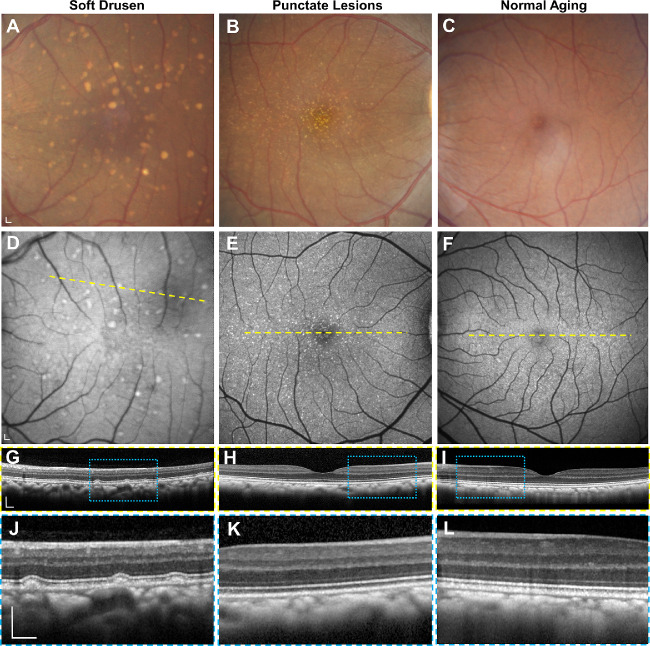
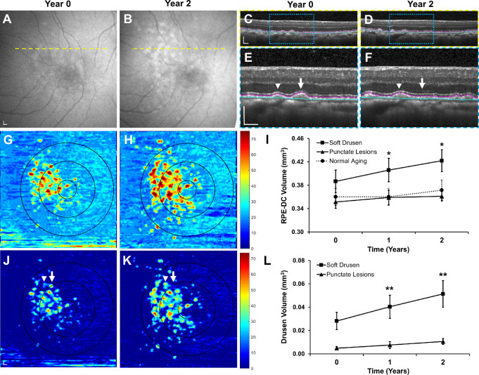
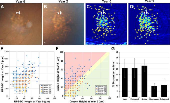
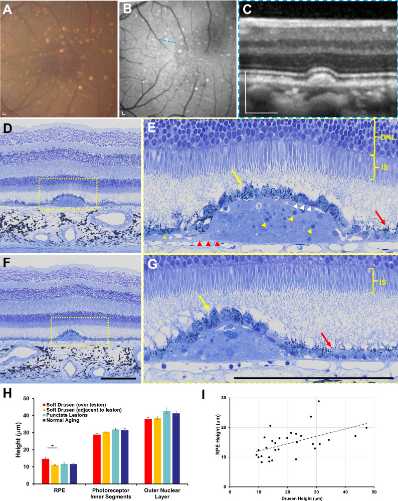
Multimodal imaging including fundus photography, spectral domain optical coherence tomography (SD-OCT), and fundus autofluorescence (FAF) were used to characterize and track individual drusen lesions in 20 aged rhesus macaques (mean age 23.3 ± 2.7 years) with drusenoid lesions over 2 years, followed by semithin histologic analysis and transmission electron microscopy (TEM).

RESULTS

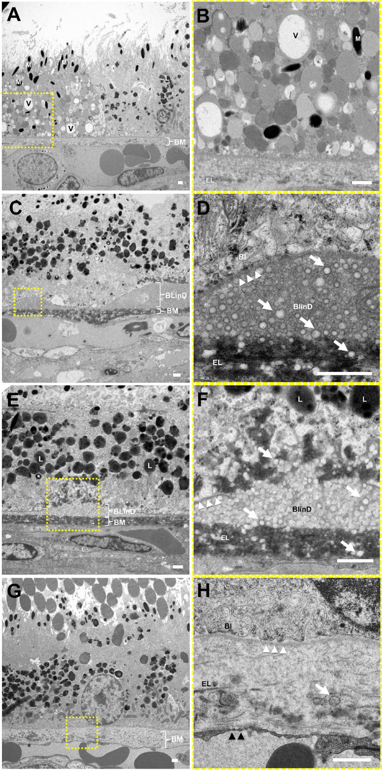
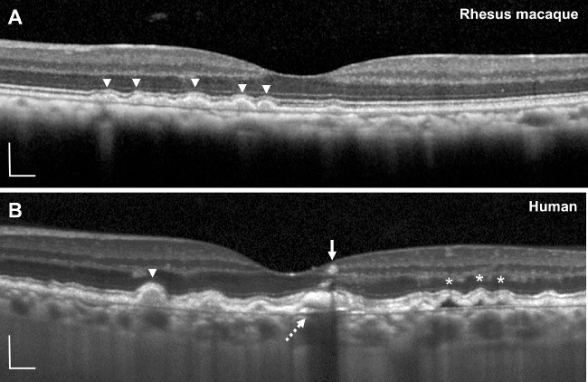
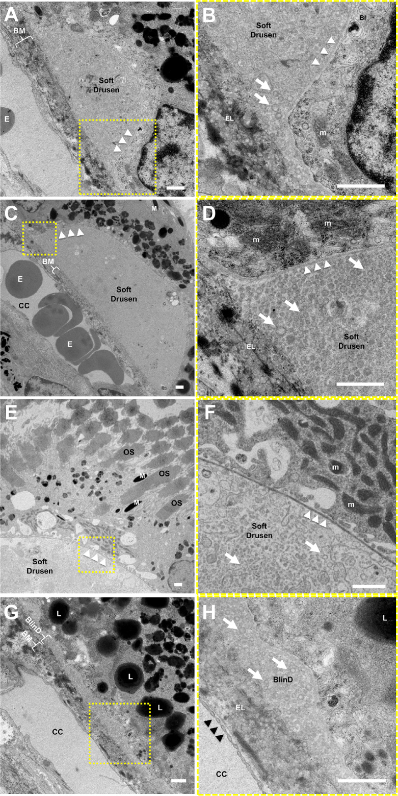
Although most drusen gradually increased in size, a portion spontaneously regressed or collapsed over 2 years. Histologic analyses showed that soft drusen exhibit hypertrophy and dysmorphia of overlying retinal pigment epithelium (RPE), as seen in early and intermediate AMD, but do not exhibit RPE atrophy, RPE migration, or photoreceptor degeneration characteristic of advanced AMD. Ultrastructure of soft drusen showed abundant lipid particles within Bruch's membrane and AMD-related basal linear deposits (BlinD) resembling those in human drusen.

CONCLUSIONS

The dynamic remodeling, histologic findings, and ultrastructural features of soft drusen in aged rhesus macaques support nonhuman primates as an animal model of early AMD and reveal important insights into drusen biogenesis and AMD development.

摘要

目的

利用活体多模态视网膜成像和离体组织学及超微结构分析,以研究恒河猴为模型的早期年龄相关性黄斑变性(AMD),对老年恒河猴软性玻璃膜疣的演变和结构进行特征描述。

方法

对 20 只年龄在 23.3 ± 2.7 岁、有软性玻璃膜疣病变超过 2 年的老年恒河猴进行多模态成像(包括眼底照相、谱域光学相干断层扫描[SD-OCT]和眼底自发荧光[FAF]),以对单个玻璃膜疣病变进行特征描述和跟踪,随后进行半薄组织学分析和透射电子显微镜(TEM)检查。

结果

尽管大多数玻璃膜疣逐渐增大,但部分在 2 年内自发消退或塌陷。组织学分析显示,软性玻璃膜疣表现为视网膜色素上皮(RPE)的过度肥大和形态异常,这与早期和中期 AMD 所见相似,但不表现为 RPE 萎缩、RPE 迁移或与晚期 AMD 相关的光感受器变性。软性玻璃膜疣的超微结构显示,Bruch 膜内有大量的脂质颗粒和 AMD 相关的基底线性沉积(BlinD),类似于人类玻璃膜疣中的情况。

结论

老年恒河猴软性玻璃膜疣的动态重塑、组织学发现和超微结构特征支持将非人类灵长类动物作为早期 AMD 的动物模型,并深入了解玻璃膜疣的发生和 AMD 的发展。

https://cdn.ncbi.nlm.nih.gov/pmc/blobs/bc9a/7326602/b2f75c0e76f7/iovs-61-2-32-f007.jpg
https://cdn.ncbi.nlm.nih.gov/pmc/blobs/bc9a/7326602/9ea9dbe1b90f/iovs-61-2-32-f001.jpg
https://cdn.ncbi.nlm.nih.gov/pmc/blobs/bc9a/7326602/77cb98e88fbf/iovs-61-2-32-f002.jpg
https://cdn.ncbi.nlm.nih.gov/pmc/blobs/bc9a/7326602/91ad1aece3b8/iovs-61-2-32-f003.jpg
https://cdn.ncbi.nlm.nih.gov/pmc/blobs/bc9a/7326602/ccb66d271088/iovs-61-2-32-f004.jpg
https://cdn.ncbi.nlm.nih.gov/pmc/blobs/bc9a/7326602/e9a644026896/iovs-61-2-32-f005.jpg
https://cdn.ncbi.nlm.nih.gov/pmc/blobs/bc9a/7326602/b55bf8f55e1b/iovs-61-2-32-f006.jpg
https://cdn.ncbi.nlm.nih.gov/pmc/blobs/bc9a/7326602/b2f75c0e76f7/iovs-61-2-32-f007.jpg

相似文献

1
Long-term Evolution and Remodeling of Soft Drusen in Rhesus Macaques.软玻璃体斑在恒河猴中的长期演变和重塑。
Invest Ophthalmol Vis Sci. 2020 Feb 7;61(2):32. doi: 10.1167/iovs.61.2.32.
2
In Vivo Multimodal Imaging of Drusenoid Lesions in Rhesus Macaques.体内灵长类猕猴脉络膜新生血管病变的多模态成像
Sci Rep. 2017 Nov 3;7(1):15013. doi: 10.1038/s41598-017-14715-z.
3
Quantitative Fundus Autofluorescence in Rhesus Macaques in Aging and Age-Related Drusen.定量眼底自发荧光在衰老和年龄相关性玻璃膜疣的恒河猴模型中的应用。
Invest Ophthalmol Vis Sci. 2020 Jul 1;61(8):16. doi: 10.1167/iovs.61.8.16.
4
Insights into pathology of cuticular drusen from integrated confocal scanning laser ophthalmoscopy imaging and corresponding spectral domain optical coherence tomography.从共焦扫描激光检眼镜成像和相应的频域光相干断层扫描中对表皮小滴的病理学认识。
Graefes Arch Clin Exp Ophthalmol. 2011 Nov;249(11):1617-25. doi: 10.1007/s00417-011-1702-0. Epub 2011 May 10.
5
Histologic and Optical Coherence Tomographic Correlates in Drusenoid Pigment Epithelium Detachment in Age-Related Macular Degeneration.年龄相关性黄斑变性中玻璃膜疣样色素上皮脱离的组织学与光学相干断层扫描相关性
Ophthalmology. 2017 May;124(5):644-656. doi: 10.1016/j.ophtha.2016.12.034. Epub 2017 Jan 30.
6
Stages of Drusen-Associated Atrophy in Age-Related Macular Degeneration Visible via Histologically Validated Fundus Autofluorescence.通过经组织学验证的眼底自发荧光观察与年龄相关性黄斑变性相关的玻璃膜疣相关萎缩的阶段。
Ophthalmol Retina. 2021 Aug;5(8):730-742. doi: 10.1016/j.oret.2020.11.006. Epub 2020 Nov 18.
7
In-vivo mapping of drusen by fundus autofluorescence and spectral-domain optical coherence tomography imaging.通过眼底自发荧光和光谱域光学相干断层扫描成像对玻璃膜疣进行体内成像。
Graefes Arch Clin Exp Ophthalmol. 2016 Jan;254(1):59-67. doi: 10.1007/s00417-015-3012-4. Epub 2015 Apr 24.
8
Calcified nodules in retinal drusen are associated with disease progression in age-related macular degeneration.视网膜玻璃膜疣中的钙化结节与年龄相关性黄斑变性的疾病进展有关。
Sci Transl Med. 2018 Nov 7;10(466). doi: 10.1126/scitranslmed.aat4544.
9
Subretinal drusenoid deposits AKA pseudodrusen.视网膜下的类脂性硬性渗出物,又名假性硬性渗出物。
Surv Ophthalmol. 2018 Nov-Dec;63(6):782-815. doi: 10.1016/j.survophthal.2018.05.005. Epub 2018 May 31.
10
Cuticular Drusen: Clinical Phenotypes and Natural History Defined Using Multimodal Imaging.角结膜皮样瘤:应用多模态成像定义的临床表型和自然史。
Ophthalmology. 2018 Jan;125(1):100-118. doi: 10.1016/j.ophtha.2017.08.033. Epub 2017 Sep 28.

引用本文的文献

1
Chromatic Pupillometry as a Putative Screening Tool for Heritable Retinal Disease in Rhesus Macaques.色觉瞳孔测量术作为恒河猴遗传性视网膜疾病的潜在筛查工具。
Transl Vis Sci Technol. 2023 Jun 1;12(6):13. doi: 10.1167/tvst.12.6.13.
2
Age-Related Macular Degeneration, a Mathematically Tractable Disease.年龄相关性黄斑变性,一种可用数学方法处理的疾病。
Invest Ophthalmol Vis Sci. 2024 Mar 5;65(3):4. doi: 10.1167/iovs.65.3.4.
3
Choroidal Changes in Rhesus Macaques in Aging and Age-Related Drusen.衰老及年龄相关性玻璃膜疣猕猴脉络膜变化

本文引用的文献

1
Vascular Response to Sildenafil Citrate in Aging and Age-Related Macular Degeneration.血管对西地那非柠檬酸酯在衰老和年龄相关性黄斑变性中的反应。
Sci Rep. 2019 Mar 25;9(1):5049. doi: 10.1038/s41598-019-41509-2.
2
Apolipoprotein A-I Mimetic Peptide L-4F Removes Bruch's Membrane Lipids in Aged Nonhuman Primates.载脂蛋白 A-I 模拟肽 L-4F 可去除老年非人灵长类动物的布鲁赫膜脂质。
Invest Ophthalmol Vis Sci. 2019 Feb 1;60(2):461-472. doi: 10.1167/iovs.18-25786.
3
A nonhuman primate model of inherited retinal disease.遗传性视网膜疾病的非人灵长类动物模型。
Invest Ophthalmol Vis Sci. 2023 Sep 1;64(12):44. doi: 10.1167/iovs.64.12.44.
4
A Spontaneous Nonhuman Primate Model of Myopic Foveoschisis.近视性黄斑劈裂的自发性非人灵长类动物模型。
Invest Ophthalmol Vis Sci. 2023 Jan 3;64(1):18. doi: 10.1167/iovs.64.1.18.
5
Relationship of Retinal Vessel Caliber with Age-Related Macular Degeneration.视网膜血管管径与年龄相关性黄斑变性的关系
J Ophthalmol. 2022 Jul 31;2022:8210599. doi: 10.1155/2022/8210599. eCollection 2022.
6
Cynomolgus monkey's choroid reference database derived from hybrid deep learning optical coherence tomography segmentation.基于混合深度学习光学相干断层扫描分割的食蟹猴脉络膜参考数据库。
Sci Rep. 2022 Aug 2;12(1):13276. doi: 10.1038/s41598-022-17699-7.
7
Tissue engineering in age-related macular degeneration: a mini-review.年龄相关性黄斑变性中的组织工程:一篇综述
J Biol Eng. 2022 May 16;16(1):11. doi: 10.1186/s13036-022-00291-y.
8
Visualizing lipid behind the retina in aging and age-related macular degeneration, via indocyanine green angiography (ASHS-LIA).通过吲哚菁绿血管造影术(ASHS-LIA)观察视网膜后脂质在衰老和年龄相关性黄斑变性中的情况。
Eye (Lond). 2022 Sep;36(9):1735-1746. doi: 10.1038/s41433-022-02016-3. Epub 2022 Mar 21.
9
A High Fat "Western-style" Diet Induces AMD-Like Features in Wildtype Mice.高脂肪“西式”饮食可诱导野生型小鼠出现 AMD 样特征。
Mol Nutr Food Res. 2022 Jun;66(11):e2100823. doi: 10.1002/mnfr.202100823. Epub 2022 Apr 28.
10
CRISPR Technology for Ocular Angiogenesis.用于眼部血管生成的CRISPR技术
Front Genome Ed. 2020 Dec 22;2:594984. doi: 10.3389/fgeed.2020.594984. eCollection 2020.
J Clin Invest. 2019 Feb 1;129(2):863-874. doi: 10.1172/JCI123980. Epub 2019 Jan 22.
4
Calcified nodules in retinal drusen are associated with disease progression in age-related macular degeneration.视网膜玻璃膜疣中的钙化结节与年龄相关性黄斑变性的疾病进展有关。
Sci Transl Med. 2018 Nov 7;10(466). doi: 10.1126/scitranslmed.aat4544.
5
Soft Drusen in Age-Related Macular Degeneration: Biology and Targeting Via the Oil Spill Strategies.年龄相关性黄斑变性中的软性玻璃疣:通过溢油策略的生物学和靶向治疗。
Invest Ophthalmol Vis Sci. 2018 Mar 20;59(4):AMD160-AMD181. doi: 10.1167/iovs.18-24882.
6
A Perspective of AMD Through the Eyes of Immunology.免疫视角下的 AMD。
Invest Ophthalmol Vis Sci. 2018 Mar 20;59(4):AMD83-AMD92. doi: 10.1167/iovs.18-23893.
7
Association Between the Cilioretinal Artery and Choroidal Neovascularization in Age-Related Macular Degeneration: A Secondary Analysis From the Age-Related Eye Disease Study.中文译文:脉络膜新生血管与年龄相关性黄斑变性的中心凹无血管区的相关性:来自年龄相关性眼病研究的二次分析。
JAMA Ophthalmol. 2018 Sep 1;136(9):1008-1014. doi: 10.1001/jamaophthalmol.2018.2650.
8
HISTOLOGY OF GEOGRAPHIC ATROPHY SECONDARY TO AGE-RELATED MACULAR DEGENERATION: A Multilayer Approach.年龄相关性黄斑变性所致地图状萎缩的组织病理学:多层分析。
Retina. 2018 Oct;38(10):1937-1953. doi: 10.1097/IAE.0000000000002182.
9
Comparison of chorioretinal layers in rhesus macaques using spectral-domain optical coherence tomography and high-resolution histological sections.应用谱域光学相干断层扫描和高分辨率组织学切片比较食蟹猴的脉络膜视网膜层。
Exp Eye Res. 2018 Mar;168:69-76. doi: 10.1016/j.exer.2018.01.012. Epub 2018 Jan 17.
10
Choroidal Changes After Suprachoroidal Injection of Triamcinolone Acetonide in Eyes With Macular Edema Secondary to Retinal Vein Occlusion.脉络膜变化后的眼 suprachoroidal 注射曲安奈德在黄斑水肿继发于视网膜静脉阻塞。
Am J Ophthalmol. 2018 Feb;186:144-151. doi: 10.1016/j.ajo.2017.11.020. Epub 2017 Dec 2.