• 文献检索
  • 文档翻译
  • 深度研究
  • 学术资讯
  • Suppr Zotero 插件Zotero 插件
  • 邀请有礼
  • 套餐&价格
  • 历史记录
应用&插件
Suppr Zotero 插件Zotero 插件浏览器插件Mac 客户端Windows 客户端微信小程序
定价
高级版会员购买积分包购买API积分包
服务
文献检索文档翻译深度研究API 文档MCP 服务
关于我们
关于 Suppr公司介绍联系我们用户协议隐私条款
关注我们

Suppr 超能文献

核心技术专利:CN118964589B侵权必究
粤ICP备2023148730 号-1Suppr @ 2026

文献检索

告别复杂PubMed语法,用中文像聊天一样搜索,搜遍4000万医学文献。AI智能推荐,让科研检索更轻松。

立即免费搜索

文件翻译

保留排版,准确专业,支持PDF/Word/PPT等文件格式,支持 12+语言互译。

免费翻译文档

深度研究

AI帮你快速写综述,25分钟生成高质量综述,智能提取关键信息,辅助科研写作。

立即免费体验

正视眼和低眼压眼中的中心凹结构与视力。

Foveal structure in nanophthalmos and visual acuity.

机构信息

Department of Ophthalmology and Visual Science, Hiroshima University Graduate School of Biomedical and Health Sciences, 1-2-3 Kasumi, Minamiku, Hiroshima, 7348551, Japan.

出版信息

Int Ophthalmol. 2021 Mar;41(3):805-813. doi: 10.1007/s10792-020-01633-9. Epub 2020 Nov 13.

DOI:10.1007/s10792-020-01633-9
PMID:33188472
原文链接:https://pmc.ncbi.nlm.nih.gov/articles/PMC7943428/
Abstract

PURPOSE

To evaluate the fovea in nanophthalmic eyes using spectral domain optical coherence tomography (SD-OCT) and OCT angiography (OCTA), and to investigate the relationship between the macular microstructure and visual acuity.

METHODS

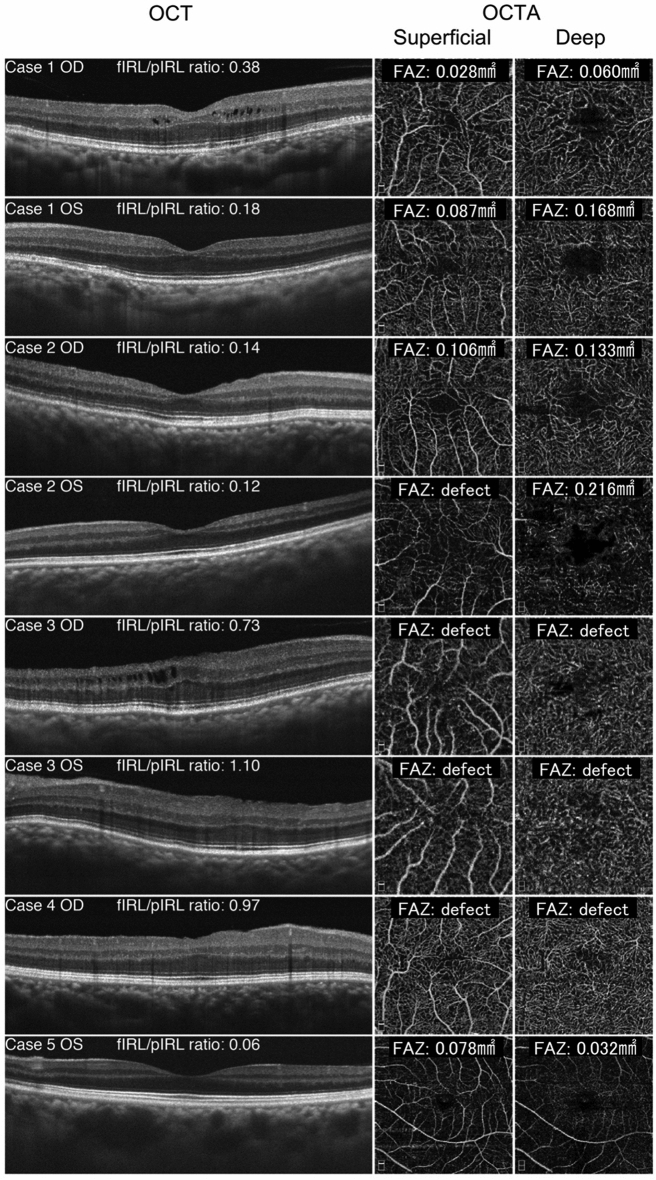
This is a retrospective case series of five nanophthalmic patients. The foveal avascular zone (FAZ) area was measured in superficial and deep vascular layers with OCTA. The thickness of the inner retinal layer (IRL) was measured with SD-OCT. The ratio of the foveal and parafoveal IRL thickness (fIRL/pIRL ratio) was calculated. The relationship between these parameters and visual acuity was then investigated.

RESULTS

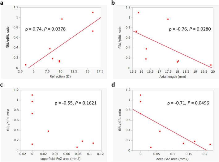
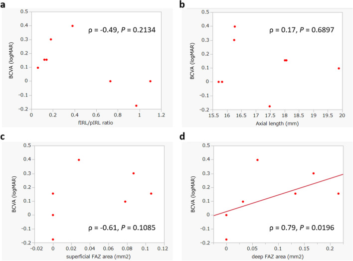
Eight eyes were identified as nanophthalmic with a mean axial length of 17.19 ± 1.44 mm (range: 15.71 to 19.88 mm). The mean best-corrected visual acuity (BCVA) in the logarithm of the minimum angle of resolution (logMAR) was 0.12 ± 0.18 (range: - 0.18 to 0.40). OCTA showed that FAZs were either absent or undeveloped in the superficial and deep capillary plexuses. Two patients did not show any visual impairments despite small FAZ and a shallow foveal depression. Although the BCVA was significantly correlated with the deep FAZ size, it did not correlate with the superficial FAZ size, axial length, or fIRL/pIRL ratio. However, the refractive error, axial length, and deep FAZ size were all significantly correlated with the fIRL/pIRL ratio.

CONCLUSIONS

The FAZs were commonly found to be small in the superficial and deep capillary plexuses. Although the deep FAZ size correlated with visual acuity, it is unclear whether the retinal microstructure and the FAZ size are responsible for the visual impairments observed in the same individuals.

摘要

目的

利用谱域光相干断层扫描(SD-OCT)和 OCT 血管造影(OCTA)评估小眼球的黄斑部,并探讨黄斑部微观结构与视力之间的关系。

方法

这是一项回顾性的小眼球病例系列研究,共纳入 5 名小眼球患者。使用 OCTA 测量浅层和深层血管层的黄斑无血管区(FAZ)面积。使用 SD-OCT 测量内视网膜层(IRL)的厚度。计算黄斑部和旁黄斑部 IRL 厚度的比值(fIRL/pIRL 比值)。然后研究这些参数与视力之间的关系。

结果

8 只眼被确定为小眼球,平均眼轴长度为 17.19±1.44mm(范围:15.71 至 19.88mm)。平均最佳矫正视力(BCVA)在最小分辨角对数(logMAR)中为 0.12±0.18(范围:-0.18 至 0.40)。OCTA 显示浅层和深层毛细血管丛中的 FAZ 要么不存在,要么发育不全。尽管 FAZ 较小且黄斑部较浅,但有 2 名患者视力不受影响。尽管 BCVA 与深层 FAZ 大小显著相关,但与浅层 FAZ 大小、眼轴长度或 fIRL/pIRL 比值均无相关性。然而,屈光不正、眼轴长度和深层 FAZ 大小均与 fIRL/pIRL 比值显著相关。

结论

浅层和深层毛细血管丛中的 FAZ 通常较小。尽管深层 FAZ 大小与视力相关,但尚不清楚视网膜微观结构和 FAZ 大小是否是导致同一患者视力下降的原因。

https://cdn.ncbi.nlm.nih.gov/pmc/blobs/8586/7943428/8905467bdea8/10792_2020_1633_Fig3_HTML.jpg
https://cdn.ncbi.nlm.nih.gov/pmc/blobs/8586/7943428/f6e608619784/10792_2020_1633_Fig1_HTML.jpg
https://cdn.ncbi.nlm.nih.gov/pmc/blobs/8586/7943428/ce006805d566/10792_2020_1633_Fig2_HTML.jpg
https://cdn.ncbi.nlm.nih.gov/pmc/blobs/8586/7943428/8905467bdea8/10792_2020_1633_Fig3_HTML.jpg
https://cdn.ncbi.nlm.nih.gov/pmc/blobs/8586/7943428/f6e608619784/10792_2020_1633_Fig1_HTML.jpg
https://cdn.ncbi.nlm.nih.gov/pmc/blobs/8586/7943428/ce006805d566/10792_2020_1633_Fig2_HTML.jpg
https://cdn.ncbi.nlm.nih.gov/pmc/blobs/8586/7943428/8905467bdea8/10792_2020_1633_Fig3_HTML.jpg

相似文献

1
Foveal structure in nanophthalmos and visual acuity.正视眼和低眼压眼中的中心凹结构与视力。
Int Ophthalmol. 2021 Mar;41(3):805-813. doi: 10.1007/s10792-020-01633-9. Epub 2020 Nov 13.
2
Unmeasurable small size superficial and deep foveal avascular zone in nanophthalmos: the Collaborative Nanophthalmos OCTA Study.无法测量的小尺寸浅层和深层中心凹无血管区在小眼球中的表现:协作性小眼球 OCTA 研究。
Br J Ophthalmol. 2019 Aug;103(8):1173-1178. doi: 10.1136/bjophthalmol-2018-312781. Epub 2018 Oct 15.
3
[Correlation of capillary plexus with visual acuity in idiopathic macular epiretinal membrane eyes using optical coherence tomography angiography].[利用光学相干断层扫描血管造影术研究特发性黄斑视网膜前膜眼中毛细血管丛与视力的相关性]
Zhonghua Yan Ke Za Zhi. 2019 Oct 11;55(10):757-762. doi: 10.3760/cma.j.issn.0412-4081.2019.10.006.
4
Foveal Hypoplasia in Patients with Stickler Syndrome.斯特奇-韦伯综合征患者的中心凹发育不良。
Ophthalmology. 2017 Jun;124(6):896-902. doi: 10.1016/j.ophtha.2017.01.046. Epub 2017 Mar 7.
5
CORRELATION OF MICROVASCULAR STRUCTURES ON OPTICAL COHERENCE TOMOGRAPHY ANGIOGRAPHY WITH VISUAL ACUITY IN RETINAL VEIN OCCLUSION.视网膜静脉阻塞患者光学相干断层扫描血管造影微血管结构与视力的相关性
Retina. 2017 Sep;37(9):1700-1709. doi: 10.1097/IAE.0000000000001403.
6
CORRELATION OF FOVEAL AVASCULAR ZONE SIZE WITH FOVEAL MORPHOLOGY IN NORMAL EYES USING OPTICAL COHERENCE TOMOGRAPHY ANGIOGRAPHY.使用光学相干断层扫描血管造影术对正常眼黄斑中心凹血管区大小与黄斑中心凹形态的相关性研究
Retina. 2015 Nov;35(11):2188-95. doi: 10.1097/IAE.0000000000000847.
7
Macular vessels density in diabetic retinopathy: quantitative assessment using optical coherence tomography angiography.糖尿病视网膜病变中的黄斑血管密度:使用光学相干断层扫描血管造影术进行定量评估。
Int Ophthalmol. 2019 Aug;39(8):1845-1859. doi: 10.1007/s10792-018-1013-0. Epub 2018 Sep 7.
8
Quantification of Diabetic Macular Ischemia Using Optical Coherence Tomography Angiography and Its Relationship with Visual Acuity.利用光相干断层扫描血管造影术对糖尿病性黄斑水肿进行定量分析及其与视力的关系。
Ophthalmology. 2017 Feb;124(2):235-244. doi: 10.1016/j.ophtha.2016.10.008. Epub 2016 Nov 23.
9
Visual Acuity and Foveal Structure in Eyes with Fragmented Foveal Avascular Zones.伴有部分性黄斑区无血管区的眼的视力和中心凹结构。
Ophthalmol Retina. 2020 May;4(5):535-544. doi: 10.1016/j.oret.2019.11.014. Epub 2019 Nov 22.
10
Optical Coherence Tomography Angiography in Central Retinal Vein Occlusion: Correlation Between the Foveal Avascular Zone and Visual Acuity.光学相干断层扫描血管造影在视网膜中央静脉阻塞中的应用:黄斑无血管区与视力的相关性
Invest Ophthalmol Vis Sci. 2016 Jul 1;57(9):OCT242-6. doi: 10.1167/iovs.15-18819.

引用本文的文献

1
Multimodal Imaging of Diabetic Retinopathy: Insights from Optical Coherence Tomography Angiography and Adaptive Optics.糖尿病视网膜病变的多模态成像:光学相干断层扫描血管造影和自适应光学的见解
Diagnostics (Basel). 2025 Jul 8;15(14):1732. doi: 10.3390/diagnostics15141732.
2
Flow and ischemic changes in retina and choroid across diabetic retinopathy spectrum: a SS-OCTA study.糖尿病视网膜病变各阶段视网膜和脉络膜的血流及缺血性改变:一项 swept-source OCTA 研究
Eye (Lond). 2025 Jun;39(8):1631-1640. doi: 10.1038/s41433-025-03639-y. Epub 2025 Feb 27.
3
Idiopathic Macular Hole Area to Foveal Avascular Zone Ratio and Its Effects on Visual Acuity Before and After Surgery.

本文引用的文献

1
Quantitative optical coherence tomography angiography of macular vascular structure and foveal avascular zone in glaucoma.青光眼黄斑血管结构及中心凹无血管区的定量光学相干断层扫描血管造影
PLoS One. 2017 Sep 21;12(9):e0184948. doi: 10.1371/journal.pone.0184948. eCollection 2017.
2
Foveal Hypoplasia in Patients with Stickler Syndrome.斯特奇-韦伯综合征患者的中心凹发育不良。
Ophthalmology. 2017 Jun;124(6):896-902. doi: 10.1016/j.ophtha.2017.01.046. Epub 2017 Mar 7.
3
Evaluation of Segmentation of the Superficial and Deep Vascular Layers of the Retina by Optical Coherence Tomography Angiography Instruments in Normal Eyes.
特发性黄斑裂孔面积与黄斑无血管区比值及其对手术前后视力的影响。
Transl Vis Sci Technol. 2025 Feb 3;14(2):22. doi: 10.1167/tvst.14.2.22.
4
Optical coherence tomography angiography in diabetic retinopathy.光学相干断层扫描血管造影在糖尿病视网膜病变中的应用。
Prog Retin Eye Res. 2023 Nov;97:101206. doi: 10.1016/j.preteyeres.2023.101206. Epub 2023 Jul 26.
5
HORNBILL: a phase I/IIa trial examining the safety, tolerability and early response of BI 764524 in patients with diabetic retinopathy and diabetic macular ischaemia-rationale, study design and protocol.犀鸟:一项 I/IIa 期临床试验,旨在研究 BI 764524 在糖尿病性视网膜病变和糖尿病性黄斑缺血患者中的安全性、耐受性和早期反应-原理、研究设计和方案。
Trials. 2022 Aug 17;23(1):669. doi: 10.1186/s13063-022-06527-y.
6
Novel ophthalmic findings and deep phenotyping in Williams-Beuren syndrome.Williams-Beuren 综合征的眼部新发现和深度表型分析。
Br J Ophthalmol. 2023 Oct;107(10):1554-1559. doi: 10.1136/bjophthalmol-2022-321103. Epub 2022 Jun 27.
7
A lightweight deep learning model for automatic segmentation and analysis of ophthalmic images.一种用于眼科图像自动分割和分析的轻量级深度学习模型。
Sci Rep. 2022 May 20;12(1):8508. doi: 10.1038/s41598-022-12486-w.
光学相干断层扫描血管造影仪对正常眼视网膜浅、深层血管层分割的评估
JAMA Ophthalmol. 2017 Mar 1;135(3):259-262. doi: 10.1001/jamaophthalmol.2016.5327.
4
Choroidal thickness findings in two siblings with nanophthalmos by swept source-OCT: a case report.经扫频源光学相干断层扫描发现的两例小眼球症同胞的脉络膜厚度:病例报告
BMC Res Notes. 2017 Jan 3;10(1):16. doi: 10.1186/s13104-016-2359-x.
5
Macular microvasculature alterations in patients with primary open-angle glaucoma: A cross-sectional study.原发性开角型青光眼患者的黄斑部微血管改变:一项横断面研究。
Medicine (Baltimore). 2016 Aug;95(33):e4341. doi: 10.1097/MD.0000000000004341.
6
Swept-Source OCT Angiography Imaging of the Foveal Avascular Zone and Macular Capillary Network Density in Diabetic Retinopathy.糖尿病视网膜病变中黄斑无血管区和黄斑毛细血管网络密度的扫频源光学相干断层扫描血管造影成像
Invest Ophthalmol Vis Sci. 2016 Jul 1;57(8):3907-13. doi: 10.1167/iovs.16-19570.
7
RETINAL AND CHOROIDAL CHANGES OF NANOPHTHALMIC EYES WITH AND WITHOUT SECONDARY GLAUCOMA.有无继发性青光眼的小眼球的视网膜和脉络膜变化
Retina. 2015 Oct;35(10):2121-9. doi: 10.1097/IAE.0000000000000564.
8
Foveal structure-function correlation in children with history of retinopathy of prematurity.有早产视网膜病变病史儿童的黄斑中心凹结构-功能相关性
Am J Ophthalmol. 2014 Sep;158(3):508-12.e2. doi: 10.1016/j.ajo.2014.05.017. Epub 2014 May 27.
9
Adaptation of the central retina for high acuity vision: cones, the fovea and the avascular zone.中央视网膜适应高敏锐度视觉:视锥细胞、中央凹和无血管区。
Prog Retin Eye Res. 2013 Jul;35:63-81. doi: 10.1016/j.preteyeres.2013.01.005. Epub 2013 Mar 15.
10
Histologic development of the human fovea from midgestation to maturity.人眼黄斑从中孕期到成熟期的组织学发育。
Am J Ophthalmol. 2012 Nov;154(5):767-778.e2. doi: 10.1016/j.ajo.2012.05.007. Epub 2012 Aug 28.