Kang Myong-Hun, Lee Sook-Jeong, Lee Min-Ho
Department of Dental Biomaterials and Institute of Biodegradable Materials, Institute of Oral Bioscience and School of Dentistry (Plus BK21 Program), Jeonbuk National University, Jeonju, Jeollabuk-do, Republic of Korea.
Department of Bioactive Material Science, Jeonbuk National University, Jeonju, Jeollabuk-do, Republic of Korea.
J Ginseng Res. 2020 Nov;44(6):823-832. doi: 10.1016/j.jgr.2020.05.003. Epub 2020 May 27.
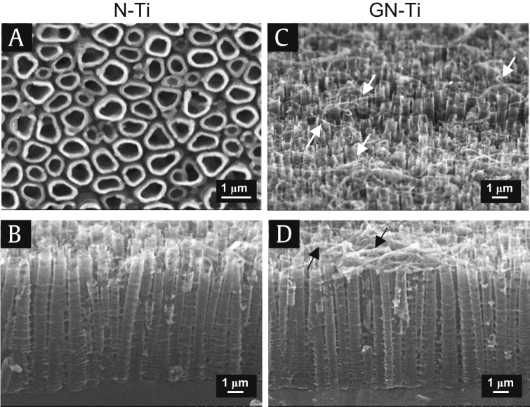
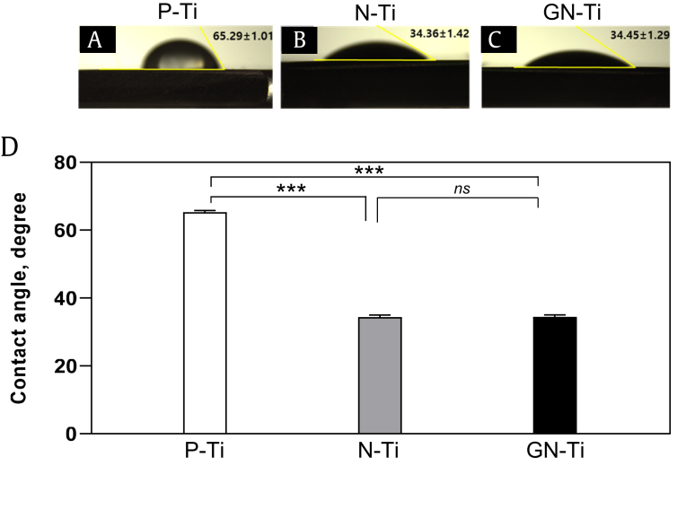
The formation of a nanotube layer on a titanium nanotube (N-Ti) plate facilitates an active reaction between bone cells and the material surface via efficient delivery of the surface materials of the dental implant into the tissues. Studies have reported that Korean Red Ginseng extracts (KRGEs) are involved in a variety of pharmacological activities: we investigated whether implantation with a KRGE-loaded N-Ti miniimplant affects osteogenesis and osseointegration.
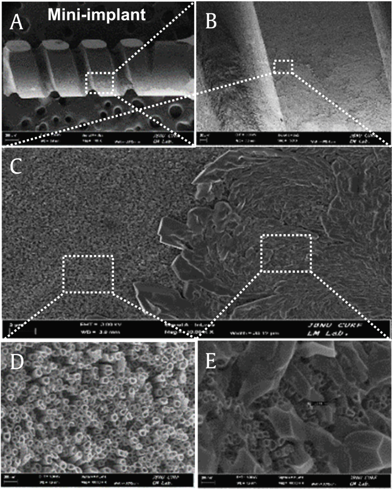
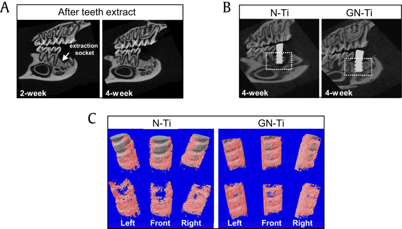
KRGE-loaded nanotubes were constructed by fabrication on pure Ti via anodization, and MC3T3-E1 cells were cultured on the N-Ti. N-Ti implants were subsequently placed on a rat's edentulous mandibular site. New bone formation and bone mineral density were measured to analyze osteogenesis and osseointegration.
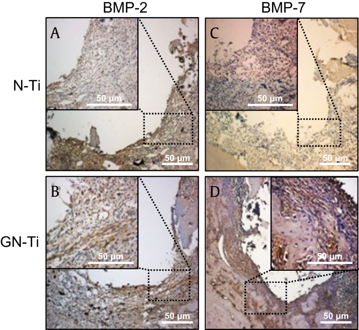
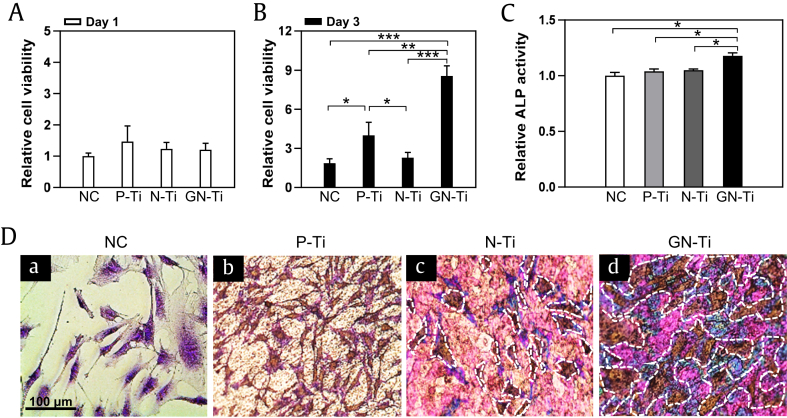
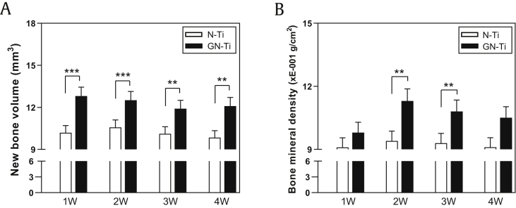
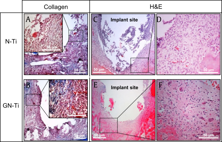
KRGE-loaded N-Ti significantly increased the proliferation and differentiation of MC3T3-E1 cells compared with cells on pure Ti without any KRGE loading. After 1-4 weeks, the periimplant tissue in the edentulous mandibular of the healed rat showed a remarkable increase in new bone formation and bone mineral density. In addition, high levels of the bone morphogenesis protein-2 and bone morphogenesis protein-7, besides collagen, were expressed in the periimplant tissues.
Our findings suggest that KRGE-induced osteogenesis and osseointegration around the miniimplant may facilitate the clinical application of dental implants.
在钛纳米管(N-Ti)板上形成纳米管层,可通过将牙种植体的表面材料有效递送至组织中,促进骨细胞与材料表面之间的活性反应。研究报道,韩国红参提取物(KRGEs)具有多种药理活性:我们研究了植入负载KRGE的N-Ti微型种植体是否会影响骨生成和骨整合。
通过阳极氧化法在纯钛上制备负载KRGE的纳米管,并将MC3T3-E1细胞培养在N-Ti上。随后将N-Ti种植体植入大鼠无牙下颌部位。测量新骨形成和骨矿物质密度,以分析骨生成和骨整合情况。
与未负载任何KRGE的纯钛上的细胞相比,负载KRGE的N-Ti显著增加了MC3T3-E1细胞的增殖和分化。1-4周后,愈合大鼠无牙下颌的种植体周围组织显示出新骨形成和骨矿物质密度显著增加。此外,种植体周围组织中除了胶原蛋白外,还表达了高水平的骨形态发生蛋白-2和骨形态发生蛋白-7。
我们的研究结果表明,KRGE诱导的微型种植体周围的骨生成和骨整合可能会促进牙种植体的临床应用。