Ramos-Gomes Fernanda, Ferreira Nathalia, Kraupner Alexander, Alves Frauke, Markus M Andrea
Translational Molecular Imaging, Max-Planck-Institute for Experimental Medicine, Göttingen, Germany.
nanoPET Pharma GmbH, Berlin, Germany.
Front Bioeng Biotechnol. 2020 Oct 30;8:588922. doi: 10.3389/fbioe.2020.588922. eCollection 2020.
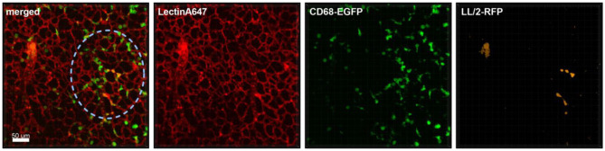
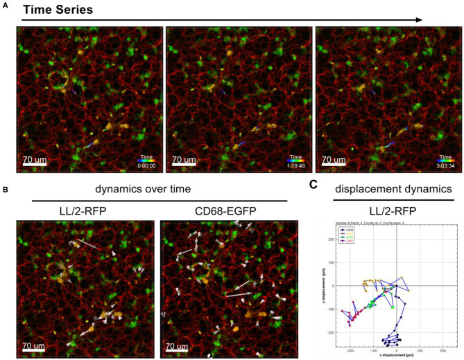
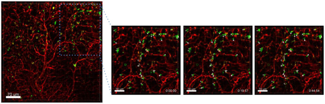
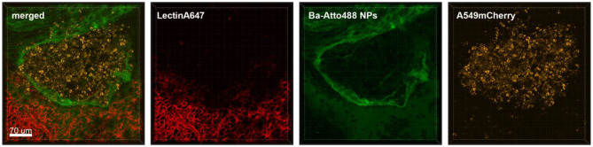
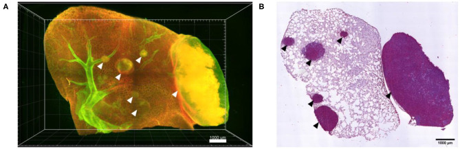
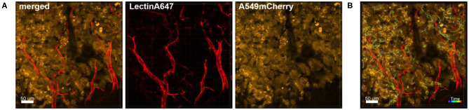
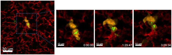
A successful clinical translation of novel nanoparticle-based cancer therapeutics requires a thorough preclinical investigation of their interaction with immune, tumor and endothelial cells as well as components of the tumor-microenvironment. Although high-resolution microscopy images of fixed tumor tissue specimens can provide valuable information in this regard, they are only static snapshots of a momentary event. Here we describe a superior alternative fluorescence microscopy approach to assess the feasibility of investigating nanoparticle-cell interactions in the mouse lung live and over time at nanometer resolution. We applied fluorescent lung tumor cells and Barium-based fluorescently labeled nanoparticles to nude mice or to CD68-EGFP transgenic mice for visualization of the monocyte-macrophage lineage. Shortly before imaging, fluorescently labeled lectin was intravenously injected for staining of the blood vessels. The lung was filled with 1% agarose and individual lung lobes were imaged over time using a confocal microscope with Airyscan technology. Time series demonstrate that live cell imaging of lung lobes can be performed for at least 4 h post mortem. Time-lapse movies illustrate the dynamics of the nanoparticles within the pulmonary circulation and their uptake by immune cells. Moreover, the exchange of nanoparticle material between cancer cells was observed over time. Fluorescent monocytes in lungs of CD68-EGFP transgenic mice could be visualized within blood vessels in the process of interaction with tumor cells and nanoparticles. This high resolution live cell imaging approach provides an excellent 4D tool to obtain valuable information on the behavior of tumor and immune cells at first encounter with nanoparticles and may contribute to the understanding of how nanoparticles interact with cells supporting the development of therapeutic strategies based on nanoparticulate drug delivery systems.
新型基于纳米颗粒的癌症治疗药物的成功临床转化需要对其与免疫细胞、肿瘤细胞、内皮细胞以及肿瘤微环境成分之间的相互作用进行全面的临床前研究。尽管固定肿瘤组织标本的高分辨率显微镜图像在这方面可以提供有价值的信息,但它们只是瞬间事件的静态快照。在此,我们描述了一种更优的荧光显微镜方法,用于评估在小鼠肺中实时且以纳米分辨率研究纳米颗粒与细胞相互作用的可行性。我们将荧光标记的肺肿瘤细胞和钡基荧光标记纳米颗粒应用于裸鼠或CD68 - EGFP转基因小鼠,以可视化单核细胞 - 巨噬细胞谱系。在成像前不久,静脉注射荧光标记的凝集素对血管进行染色。肺用1%的琼脂糖填充,使用配备Airyscan技术的共聚焦显微镜对各个肺叶进行实时成像。时间序列表明,肺叶的活细胞成像可以在死后至少进行4小时。延时电影展示了纳米颗粒在肺循环中的动态以及它们被免疫细胞摄取的过程。此外,随着时间的推移,观察到癌细胞之间纳米颗粒物质的交换。在CD68 - EGFP转基因小鼠肺中的荧光单核细胞在与肿瘤细胞和纳米颗粒相互作用的过程中可在血管内可视化。这种高分辨率活细胞成像方法提供了一个出色的四维工具,可获取肿瘤细胞和免疫细胞首次接触纳米颗粒时行为的有价值信息,并可能有助于理解纳米颗粒如何与细胞相互作用,从而支持基于纳米颗粒药物递送系统的治疗策略的开发。