Department of Pathology and Laboratory Medicine, Atlanta, GA, USA.
Department of Bioinformatics, Emory University, Atlanta, GA, USA.
Histopathology. 2021 May;78(6):791-804. doi: 10.1111/his.14304. Epub 2021 Mar 8.
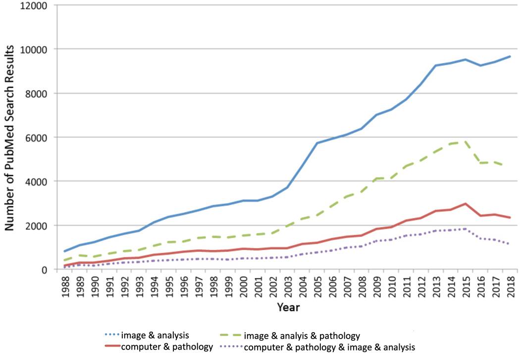
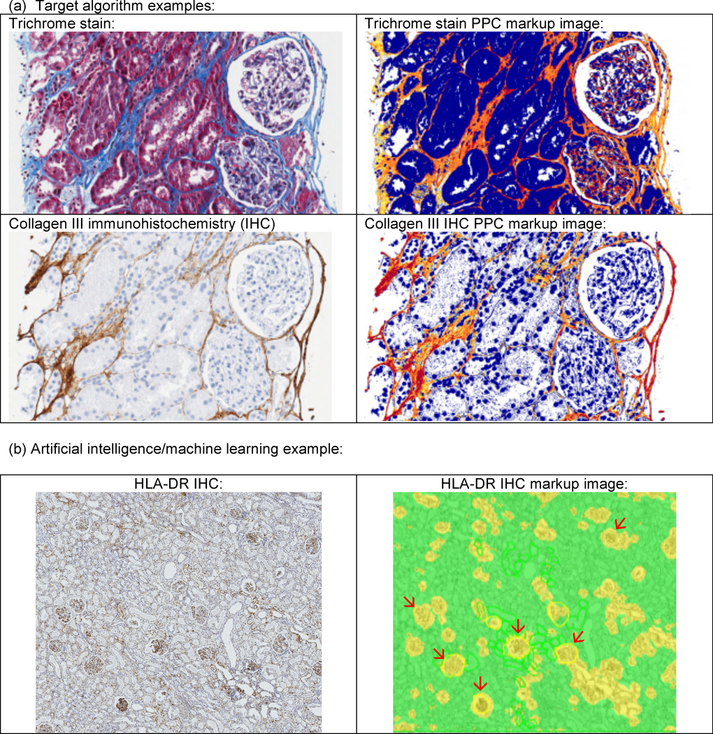
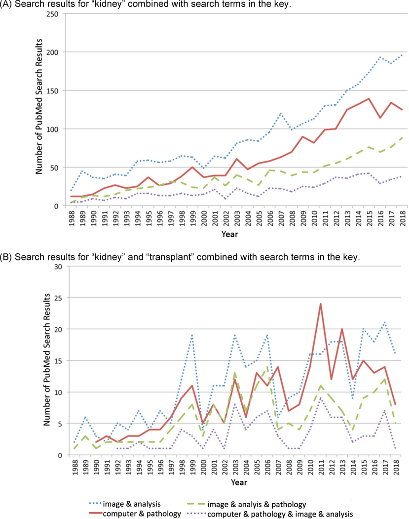
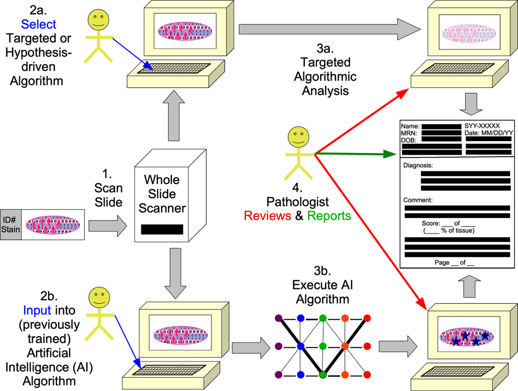
Whole slide imaging, which is an important technique in the field of digital pathology, has recently been the subject of increased interest and avenues for utilisation, and with more widespread whole slide image (WSI) utilisation, there will also be increased interest in and implementation of image analysis (IA) techniques. IA includes artificial intelligence (AI) and targeted or hypothesis-driven algorithms. In the overall pathology field, the number of citations related to these topics has increased in recent years. Renal pathology is one anatomical pathology subspecialty that has utilised WSIs and IA algorithms; it can be argued that renal transplant pathology could be particularly suited for whole slide imaging and IA, as renal transplant pathology is frequently classified by use of the semiquantitative Banff classification of renal allograft pathology. Hypothesis-driven/targeted algorithms have been used in the past for the assessment of a variety of features in the kidney (e.g. interstitial fibrosis, tubular atrophy, inflammation); in recent years, the amount of research has particularly increased in the area of AI/machine learning for the identification of glomeruli, for histological segmentation, and for other applications. Deep learning is the form of machine learning that is most often used for such AI approaches to the 'big data' of pathology WSIs, and deep learning methods such as artificial neural networks (ANNs)/convolutional neural networks (CNNs) are utilised. Unsupervised and supervised AI algorithms can be employed to accomplish image or semantic classification. In this review, AI and other IA algorithms applied to WSIs are discussed, and examples from renal pathology are covered, with an emphasis on renal transplant pathology.
全切片成像,作为数字病理学领域的一项重要技术,近来受到了越来越多的关注,并为其应用开辟了新途径。随着全切片图像(WSI)的更广泛应用,人们对图像分析(IA)技术的兴趣也将增加并得到实施。IA 包括人工智能(AI)和有针对性或基于假设的算法。在整个病理学领域,近年来与这些主题相关的引用数量有所增加。肾脏病理学是一个利用 WSI 和 IA 算法的解剖病理学亚专科;可以说,肾脏移植病理学特别适合全切片成像和 IA,因为肾脏移植病理学通常使用半定量 Banff 肾脏移植病理学分类来进行分类。过去,已经使用基于假设/有针对性的算法来评估肾脏的各种特征(例如间质纤维化、肾小管萎缩、炎症);近年来,人工智能/机器学习在识别肾小球、组织学分割和其他应用领域的研究特别增加。深度学习是最常用于此类 AI 方法处理病理学 WSI“大数据”的机器学习形式,并且使用了诸如人工神经网络(ANN)/卷积神经网络(CNN)等深度学习方法。可以使用无监督和监督 AI 算法来实现图像或语义分类。在这篇综述中,讨论了应用于 WSI 的 AI 和其他 IA 算法,并介绍了肾脏病理学的示例,重点是肾脏移植病理学。