The Lydia Becker Institute of Immunology and Inflammation, Faculty of Biology, Medicine and Health, University of Manchester, Manchester, United Kingdom.
Department of Cardiothoracic Surgery, Manchester University NHS Foundation Trust, Manchester, United Kingdom.
Biophys J. 2020 Dec 15;119(12):2403-2417. doi: 10.1016/j.bpj.2020.10.041. Epub 2020 Nov 18.
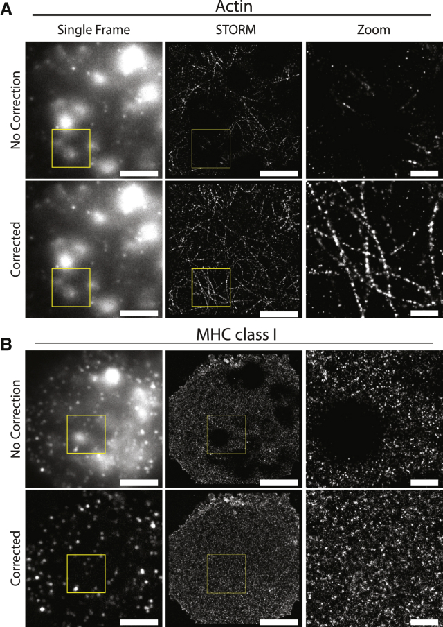
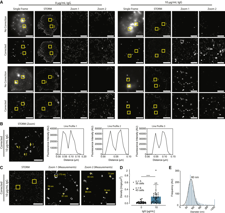
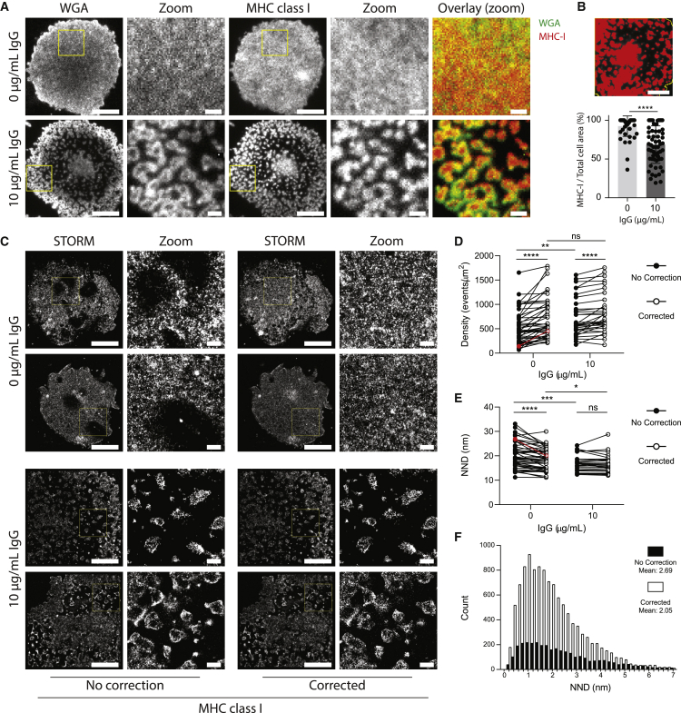
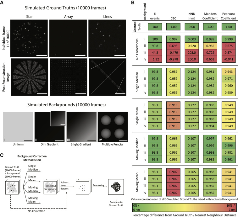
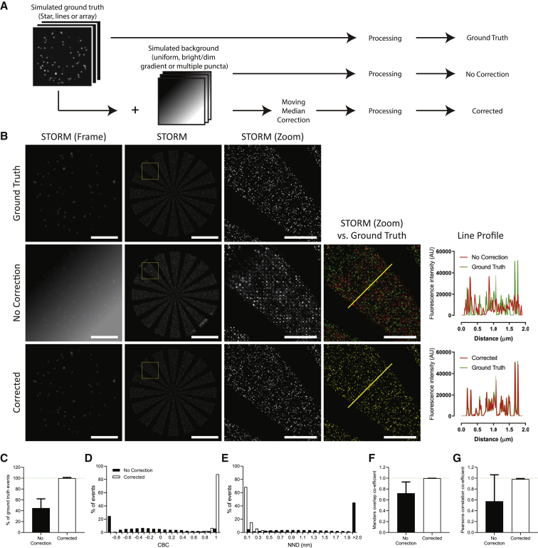
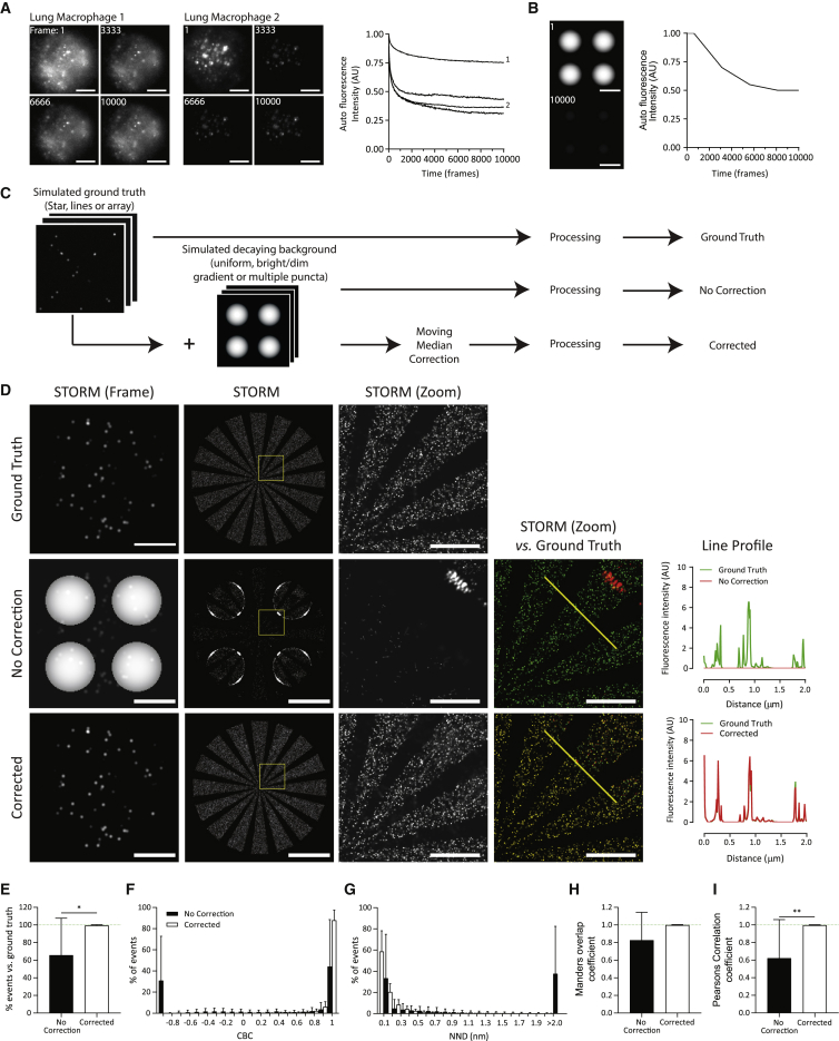
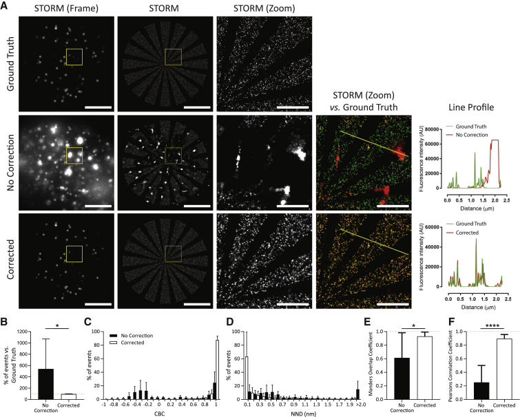
Observing the cell surface and underlying cytoskeleton at nanoscale resolution using super-resolution microscopy has enabled many insights into cell signaling and function. However, the nanoscale dynamics of tissue-specific immune cells have been relatively little studied. Tissue macrophages, for example, are highly autofluorescent, severely limiting the utility of light microscopy. Here, we report a correction technique to remove autofluorescent noise from stochastic optical reconstruction microscopy (STORM) data sets. Simulations and analysis of experimental data identified a moving median filter as an accurate and robust correction technique, which is widely applicable across challenging biological samples. Here, we used this method to visualize lung macrophages activated through Fc receptors by antibody-coated glass slides. Accurate, nanoscale quantification of macrophage morphology revealed that activation induced the formation of cellular protrusions tipped with MHC class I protein. These data are consistent with a role for lung macrophage protrusions in antigen presentation. Moreover, the tetraspanin protein CD81, known to mark extracellular vesicles, appeared in ring-shaped structures (mean diameter 93 ± 50 nm) at the surface of activated lung macrophages. Thus, a moving median filter correction technique allowed us to quantitatively analyze extracellular secretions and membrane structure in tissue-derived immune cells.
使用超分辨率显微镜观察细胞表面和底层细胞骨架,可以深入了解细胞信号转导和功能。然而,组织特异性免疫细胞的纳米级动力学研究相对较少。例如,组织巨噬细胞的自体荧光性很强,严重限制了光学显微镜的应用。在这里,我们报告了一种从随机光学重建显微镜(STORM)数据集去除自体荧光噪声的校正技术。模拟和实验数据分析确定了移动中位数滤波器是一种准确且稳健的校正技术,广泛适用于具有挑战性的生物样本。在这里,我们使用这种方法来可视化通过抗体包被的载玻片激活的肺巨噬细胞。对巨噬细胞形态的精确纳米级定量分析表明,激活诱导了带有 MHC Ⅰ类蛋白的细胞突起的形成。这些数据与肺巨噬细胞突起在抗原呈递中的作用一致。此外,已知标记细胞外囊泡的四跨膜蛋白 CD81 出现在激活的肺巨噬细胞表面的环形结构中(平均直径 93 ± 50nm)。因此,移动中位数滤波器校正技术使我们能够定量分析组织来源免疫细胞中的细胞外分泌和膜结构。