National Cancer Institute, Center for Cancer Research, Laboratory of Receptor Biology and Gene Expression, Bethesda, MD, United States.
J Mol Biol. 2021 Mar 19;433(6):166720. doi: 10.1016/j.jmb.2020.11.019. Epub 2020 Nov 20.
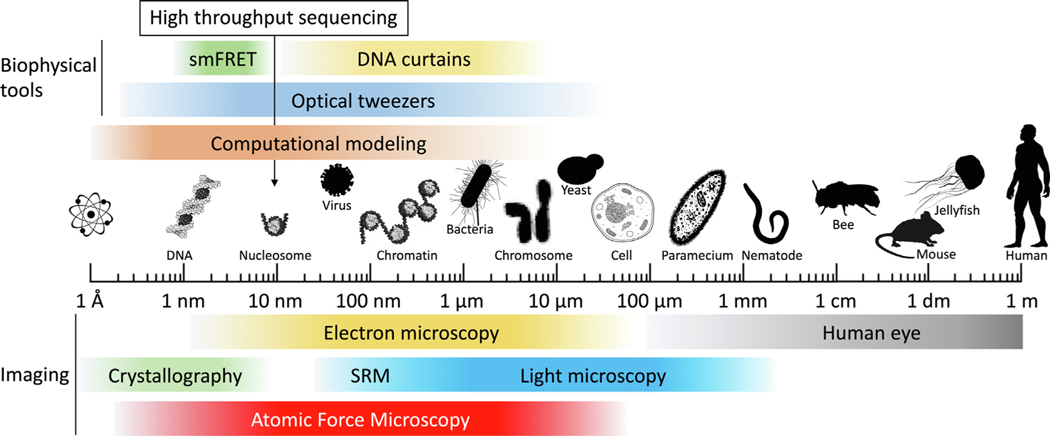
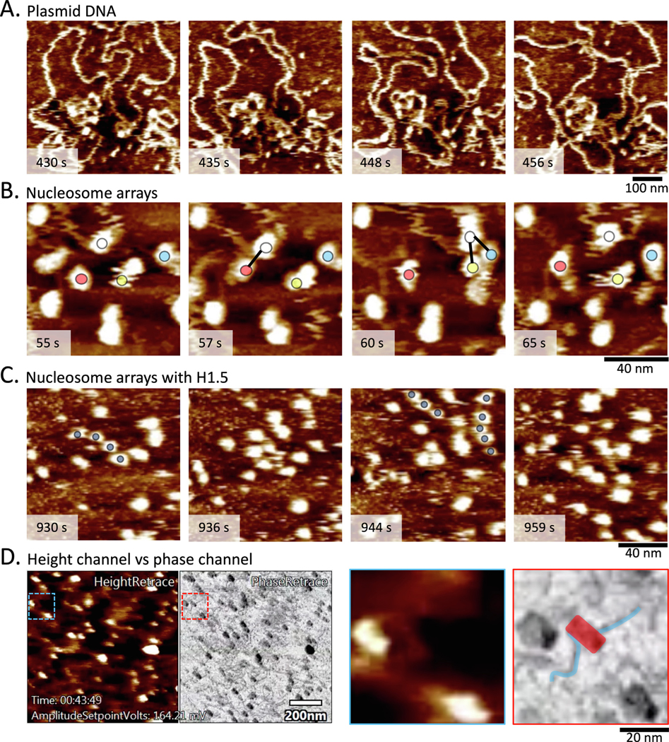
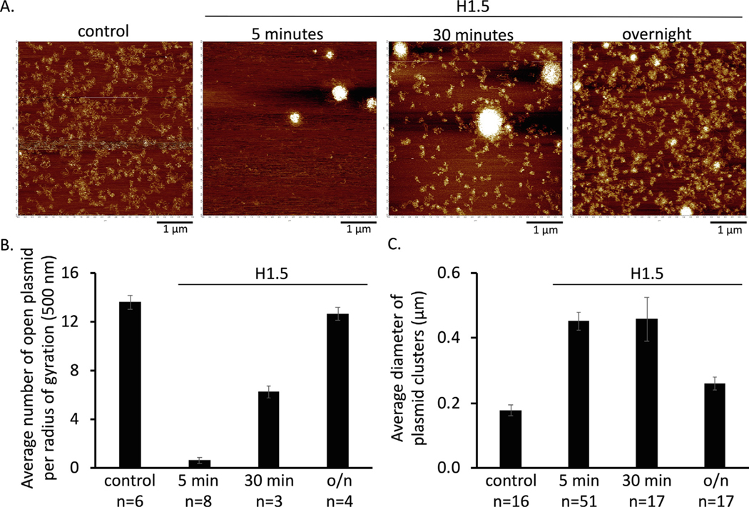
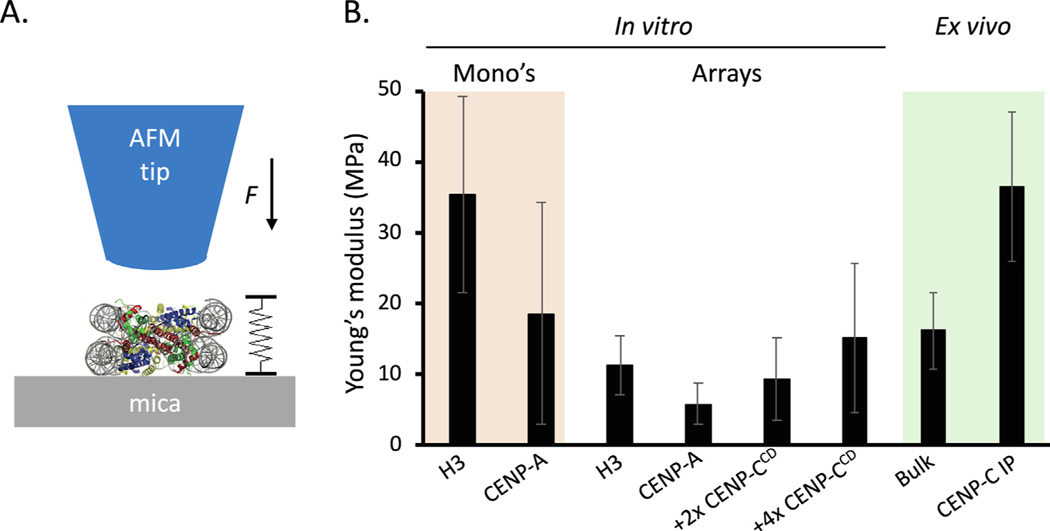
Chromatin is the epigenomic platform for diverse nuclear processes such as DNA repair, replication, transcription, telomere, and centromere function. In cancer cells, mutations in key processes result in DNA amplification, chromosome translocations, and chromothripsis, severely distorting the natural chromatin state. In normal and diseased states, dozens of chromatin effectors alter the physical integrity and dynamics of chromatin at the level of both single nucleosomes and arrays of nucleosomes folded into 3-dimensional shapes. Integrating these length scales, from the 10 nm sized nucleosome to mitotic chromosomes, whilst jostling within the crowded environment of the cell, cannot yet be achieved by a single technology. In this review, we discuss tools that have proven powerful in the investigation of nucleosome and chromatin fiber dynamics. We also provide a deeper focus into atomic force microscopy (AFM) applications that can bridge diverse length and time scales. Using time course AFM, we observe that chromatin condensation by H1.5 is dynamic, whereas using nano-indentation force spectroscopy we observe that both histone variants and nucleosome binding partners alter material properties of individual nucleosomes. Finally, we demonstrate how high-speed AFM can visualize plasmid DNA dynamics, intermittent nucleosome-nucleosome contacts, and changes in nucleosome phasing along a contiguous chromatin fiber. Altogether, the development of innovative technologies holds the promise of revealing the secret lives of nucleosomes, potentially bridging the gaps in our understanding of how chromatin works within living cells and tissues.
染色质是多种核过程的表观基因组平台,如 DNA 修复、复制、转录、端粒和着丝粒功能。在癌细胞中,关键过程的突变导致 DNA 扩增、染色体易位和染色质碎裂,严重扭曲了自然染色质状态。在正常和患病状态下,数十种染色质效应物改变单个核小体和折叠成三维形状的核小体阵列的物理完整性和动力学。整合这些长度尺度,从 10nm 大小的核小体到有丝分裂染色体,同时在细胞拥挤的环境中推挤,目前还不能仅通过单一技术实现。在这篇综述中,我们讨论了已被证明在研究核小体和染色质纤维动力学方面非常有效的工具。我们还更深入地探讨了原子力显微镜(AFM)在跨越不同长度和时间尺度方面的应用。使用时程 AFM,我们观察到 H1.5 引起的染色质浓缩是动态的,而使用纳米压痕力谱学,我们观察到组蛋白变体和核小体结合伴侣都改变了单个核小体的材料特性。最后,我们展示了高速 AFM 如何可视化质粒 DNA 动力学、间歇性核小体-核小体接触以及沿连续染色质纤维的核小体相位变化。总之,创新技术的发展有望揭示核小体的秘密生活,有可能弥合我们对染色质在活细胞和组织中如何工作的理解中的差距。