Beheshti Iman, Sone Daichi, Maikusa Norihide, Kimura Yukio, Shigemoto Yoko, Sato Noriko, Matsuda Hiroshi
Department of Human Anatomy and Cell Science, Rady Faculty of Health Sciences, Max Rady College of Medicine, University of Manitoba, Winnipeg, MB, Canada.
Cyclotron and Drug Discovery Research Center, Southern Tohoku Research Institute for Neuroscience, Koriyama, Japan.
Front Neurol. 2020 Nov 3;11:580713. doi: 10.3389/fneur.2020.580713. eCollection 2020.
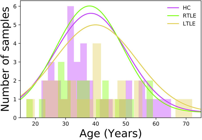
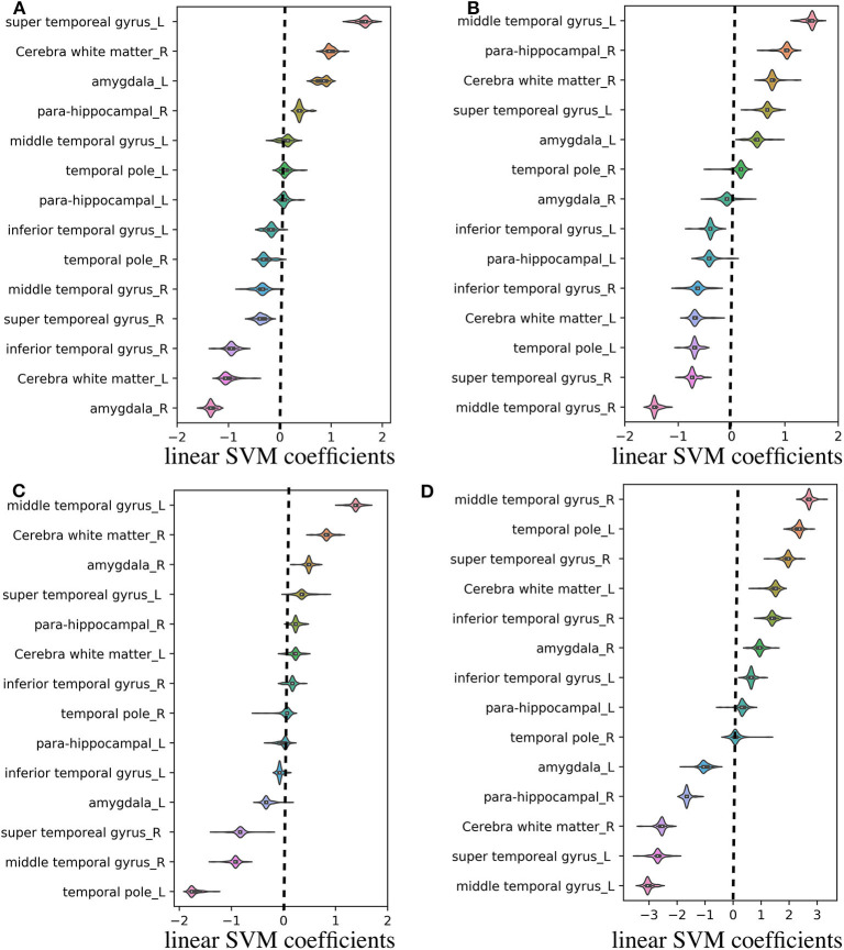
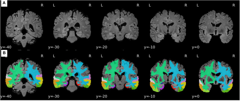
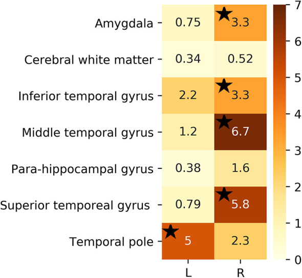
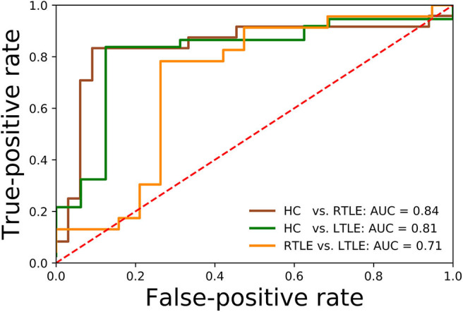
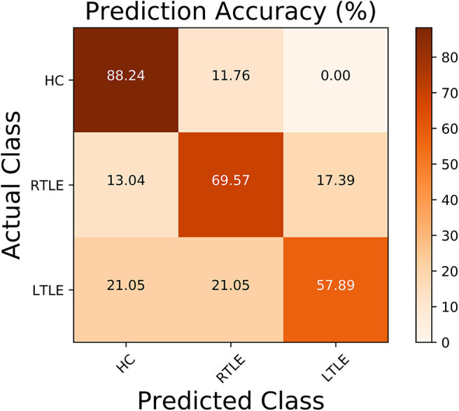
In this study, we investigated the ability of fluid-attenuated inversion recovery (FLAIR) data coupled with machine-leaning algorithms to differentiate normal and epileptic brains and identify the laterality of focus side in temporal lobe epilepsy (TLE) patients with visually negative MRI. The MRI data were acquired on a 3-T MR system (Philips Medical Systems). After pre-proceeding stage, the FLAIR signal intensities were extracted from specific regions of interest, such as the amygdala, cerebral white matter, inferior temporal gyrus, middle temporal gyrus, parahippocampal gyrus, superior temporal gyrus, and temporal pole, and fed into a classification framework followed by a support vector machine as classifier. The proposed lateralization framework was assessed in a group of MRI-negative unilateral TLE patients ( = 42; 23 left TLE and 19 right TLE) and 34 healthy controls (HCs) based on a leave-one-out cross-validation strategy. Using the FLAIR data, we obtained a 75% accuracy for discriminating the three groups, as well as 87.71, 83.01, and 76.19% accuracies for HC/right TLE, HC/left TLE, and left TLE/right TLE tasks, respectively. The experimental results show that FLAIR data can potentially be considered an informative biomarker for improving the pre-surgical diagnostic confidence in patients with MRI-negative TLE.
在本研究中,我们调查了液体衰减反转恢复(FLAIR)数据结合机器学习算法区分正常大脑和癫痫大脑以及识别视觉上MRI阴性的颞叶癫痫(TLE)患者病灶侧别(患侧)的能力。MRI数据是在一台3-T MR系统(飞利浦医疗系统公司)上采集的。在预处理阶段之后,从特定感兴趣区域提取FLAIR信号强度,这些区域包括杏仁核、脑白质、颞下回、颞中回、海马旁回、颞上回和颞极,并将其输入到一个分类框架中,随后使用支持向量机作为分类器。基于留一法交叉验证策略,在一组MRI阴性的单侧TLE患者(n = 42;23例左侧TLE和19例右侧TLE)和34名健康对照者(HCs)中评估所提出的侧别化框架。利用FLAIR数据,我们在区分这三组时获得了75%的准确率,在HC/右侧TLE、HC/左侧TLE和左侧TLE/右侧TLE任务中分别获得了87.71%、83.01%和76.19%的准确率。实验结果表明,FLAIR数据有可能被视为一种信息性生物标志物,用于提高MRI阴性TLE患者术前诊断的信心。