• 文献检索
  • 文档翻译
  • 深度研究
  • 学术资讯
  • Suppr Zotero 插件Zotero 插件
  • 邀请有礼
  • 套餐&价格
  • 历史记录
应用&插件
Suppr Zotero 插件Zotero 插件浏览器插件Mac 客户端Windows 客户端微信小程序
定价
高级版会员购买积分包购买API积分包
服务
文献检索文档翻译深度研究API 文档MCP 服务
关于我们
关于 Suppr公司介绍联系我们用户协议隐私条款
关注我们

Suppr 超能文献

核心技术专利:CN118964589B侵权必究
粤ICP备2023148730 号-1Suppr @ 2026

文献检索

告别复杂PubMed语法,用中文像聊天一样搜索,搜遍4000万医学文献。AI智能推荐,让科研检索更轻松。

立即免费搜索

文件翻译

保留排版,准确专业,支持PDF/Word/PPT等文件格式,支持 12+语言互译。

免费翻译文档

深度研究

AI帮你快速写综述,25分钟生成高质量综述,智能提取关键信息,辅助科研写作。

立即免费体验

使用锥形束计算机断层扫描对牙髓显微外科术后进行的一年期影像学愈合评估。

A One-Year Radiographic Healing Assessment after Endodontic Microsurgery Using Cone-Beam Computed Tomographic Scans.

作者信息

Kang Sumi, Ha Se-Won, Kim Ukseong, Kim Sunil, Kim Euiseong

机构信息

Microscope Center, Department of Conservative Dentistry and Oral Science Research Center, College of Dentistry, Yonsei University, Seoul 03722, Korea.

出版信息

J Clin Med. 2020 Nov 19;9(11):3714. doi: 10.3390/jcm9113714.

DOI:10.3390/jcm9113714
PMID:33228002
原文链接:https://pmc.ncbi.nlm.nih.gov/articles/PMC7699244/
Abstract

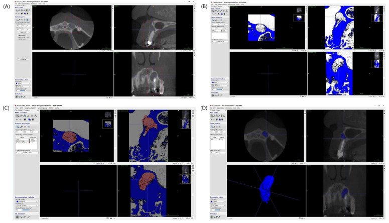
This study aimed to evaluate one-year radiographic healing after endodontic microsurgery using CBCT with modified PENN 3D criteria and to compare the outcome with results evaluated using Molven's criteria. A total of 107 teeth from 96 patients were evaluated one year after endodontic microsurgery by using CBCT scans with modified PENN 3D criteria and periapical radiographs with Molven's criteria. Both preoperative and postoperative lesion volumes were calculated using ITK-SNAP (free software). Radiographic healing assessment using periapical radiographs and CBCT images, and preoperative and postoperative lesion volume measurements were performed independently by two examiners. The assessment using Molven's criteria resulted in 75 complete healings, 18 incomplete healings, eight uncertain healings, and six unsatisfactory healings. Based on modified PENN 3D criteria, 64 teeth were categorized as complete healing, 29 teeth as limited healing, six teeth as uncertain healing, and eight teeth as unsatisfactory healing. With the one-year follow-up, CBCT scans showed a lower healing tendency than did periapical radiography. The volumes of apical radiolucency after the surgery were reduced by 77.7% on average at one-year follow up.

摘要

本研究旨在使用改良的PENN 3D标准通过CBCT评估根管显微外科手术后一年的影像学愈合情况,并将结果与使用Molven标准评估的结果进行比较。对96例患者的107颗牙齿在根管显微外科手术后一年进行评估,使用改良的PENN 3D标准进行CBCT扫描,并使用Molven标准进行根尖片检查。术前和术后的病变体积均使用ITK-SNAP(免费软件)进行计算。根尖片和CBCT图像的影像学愈合评估以及术前和术后病变体积测量由两名检查者独立进行。使用Molven标准评估的结果为75例完全愈合、18例不完全愈合、8例愈合情况不确定和6例愈合情况不满意。基于改良的PENN 3D标准,64颗牙齿被归类为完全愈合,29颗牙齿为有限愈合,6颗牙齿为愈合情况不确定,8颗牙齿为愈合情况不满意。经过一年的随访,CBCT扫描显示的愈合趋势低于根尖片检查。在一年的随访中,手术后根尖透影区的体积平均减少了77.7%。

https://cdn.ncbi.nlm.nih.gov/pmc/blobs/8f44/7699244/14c19c45134d/jcm-09-03714-g002.jpg
https://cdn.ncbi.nlm.nih.gov/pmc/blobs/8f44/7699244/25f25340a9ad/jcm-09-03714-g001.jpg
https://cdn.ncbi.nlm.nih.gov/pmc/blobs/8f44/7699244/14c19c45134d/jcm-09-03714-g002.jpg
https://cdn.ncbi.nlm.nih.gov/pmc/blobs/8f44/7699244/25f25340a9ad/jcm-09-03714-g001.jpg
https://cdn.ncbi.nlm.nih.gov/pmc/blobs/8f44/7699244/14c19c45134d/jcm-09-03714-g002.jpg

相似文献

1
A One-Year Radiographic Healing Assessment after Endodontic Microsurgery Using Cone-Beam Computed Tomographic Scans.使用锥形束计算机断层扫描对牙髓显微外科术后进行的一年期影像学愈合评估。
J Clin Med. 2020 Nov 19;9(11):3714. doi: 10.3390/jcm9113714.
2
A Comparison of 2- and 3-dimensional Healing Assessment after Endodontic Surgery Using Cone-beam Computed Tomographic Volumes or Periapical Radiographs.根管手术后使用锥形束 CT 容积或根尖片评估 2 维和 3 维愈合的比较。
J Endod. 2017 Jul;43(7):1072-1079. doi: 10.1016/j.joen.2017.02.007. Epub 2017 May 17.
3
Two- and three-dimensional healing assessment after endodontic microsurgery in through-and-through periapical lesions: 5-year follow-up from a randomized controlled trial.经根管显微外科治疗贯穿性根尖周病变的二维和三维愈合评估:一项随机对照试验的 5 年随访。
Int Endod J. 2024 Sep;57(9):1180-1199. doi: 10.1111/iej.14084. Epub 2024 May 17.
4
Patient-centric outcome assessment of endodontic microsurgery using periapical radiography versus cone beam computed tomography: A randomized clinical trial.采用根尖放射线摄影术与锥形束计算机断层摄影术评估牙髓微创手术的以患者为中心的结局:一项随机临床试验。
Int Endod J. 2023 Jan;56(1):3-16. doi: 10.1111/iej.13837. Epub 2022 Oct 2.
5
Clinical Outcome and Predictors of Endodontic Microsurgery Using Cone-beam Computed Tomography: A Retrospective Cohort Study.使用锥形束计算机断层扫描的根管显微镜手术的临床结果和预测因素:一项回顾性队列研究。
J Endod. 2023 Nov;49(11):1464-1471. doi: 10.1016/j.joen.2023.08.011. Epub 2023 Aug 24.
6
Efficacy of mixture of injectable-platelet-rich fibrin and type-1 collagen particles on the closure of through-and-through periapical bone defects: A randomized controlled trial.注射用富血小板纤维蛋白与I型胶原颗粒混合物对贯通性根尖骨缺损封闭的疗效:一项随机对照试验。
Int Endod J. 2023 Oct;56(10):1197-1211. doi: 10.1111/iej.13954. Epub 2023 Jul 17.
7
Periapical healing following endodontic microsurgery with collagen-based bone-filling material: A randomized controlled clinical trial.根管显微手术应用基于胶原蛋白的骨填充材料后的根尖周愈合:一项随机对照临床试验。
Int Endod J. 2023 Dec;56(12):1446-1458. doi: 10.1111/iej.13973. Epub 2023 Sep 11.
8
2D and 3D radiographic outcome assessment of the effect of guided tissue regeneration using resorbable collagen membrane in the healing of through-and-through periapical lesions - a randomized controlled trial.采用可吸收胶原膜引导组织再生治疗贯穿性根尖周病变愈合的二维和三维放射学评估——一项随机对照试验。
Int Endod J. 2019 Jul;52(7):935-948. doi: 10.1111/iej.13098. Epub 2019 Mar 6.
9
Impact of Platelet-rich Plasma in the Healing of Through-and-through Periapical Lesions Using 2-dimensional and 3-dimensional Evaluation: A Randomized Controlled Trial.富血小板血浆对经皮根尖周病变愈合的影响:二维和三维评估的随机对照试验。
J Endod. 2020 Sep;46(9):1167-1184. doi: 10.1016/j.joen.2020.06.004. Epub 2020 Jun 15.
10
Clinical and radiological outcomes of dynamic navigation in endodontic microsurgery: a prospective study.牙髓显微外科中动态导航的临床和放射学效果:一项前瞻性研究。
Clin Oral Investig. 2023 Sep;27(9):5317-5329. doi: 10.1007/s00784-023-05152-6. Epub 2023 Aug 2.

引用本文的文献

1
Assessment of the occurrence of apical periodontitis and endodontically treated/non-treated teeth in a Lower Austrian patient population treated for osteoporosis: a cohort study.对奥地利下奥地利州接受骨质疏松症治疗的患者群体中根尖周炎及根管治疗/未治疗牙齿发生率的评估:一项队列研究。
Ann Med Surg (Lond). 2024 Aug 14;86(9):5049-5057. doi: 10.1097/MS9.0000000000002443. eCollection 2024 Sep.
2
The use of guided tissue regeneration in endodontic Microsurgery: Setting a threshold.引导组织再生技术在牙髓显微外科中的应用:设定一个阈值。
Saudi Dent J. 2024 Mar;36(3):461-465. doi: 10.1016/j.sdentj.2023.12.005. Epub 2023 Dec 12.
3

本文引用的文献

1
3D Assessment of Endodontic Lesions with a Low-Dose CBCT Protocol.采用低剂量锥形束计算机断层扫描方案对牙髓病变进行三维评估。
Dent J (Basel). 2020 May 13;8(2):51. doi: 10.3390/dj8020051.
2
Radiographic Assessment of Bone Healing Using Cone-beam Computed Tomographic Scans 1 and 5 Years after Apical Surgery.使用锥形束计算机断层扫描评估根尖手术后 1 年和 5 年的骨愈合情况。
J Endod. 2019 Nov;45(11):1307-1313. doi: 10.1016/j.joen.2019.08.008. Epub 2019 Sep 19.
3
A 10-year Follow-up Study of 119 Teeth Treated with Apical Surgery and Root-end Filling with Mineral Trioxide Aggregate.
Application of advanced platelet-rich fibrin for through-and-through bony defect during endodontic surgery: Three case reports and review of the literature.
富血小板纤维蛋白在牙髓外科贯通性骨缺损中的应用:三例病例报告及文献综述
World J Clin Cases. 2023 Jun 16;11(17):4168-4178. doi: 10.12998/wjcc.v11.i17.4168.
4
Volumetric Assessment of Apical Periodontitis Using Cone-Beam Computed Tomography-A Systematic Review.使用锥形束计算机断层扫描评估根尖周炎的容积评估——系统评价。
Int J Environ Res Public Health. 2023 Feb 8;20(4):2940. doi: 10.3390/ijerph20042940.
5
Outcomes of endodontic microsurgery using different calcium silicate-based retrograde filling materials: a cohort retrospective cone-beam computed tomographic analysis.不同硅酸钙基逆行充填材料在根管显微外科中的应用效果:一项基于锥形束 CT 的回顾性队列研究。
BMC Oral Health. 2023 Feb 3;23(1):70. doi: 10.1186/s12903-023-02782-w.
6
Plasma Rich in Growth Factors in the Treatment of Endodontic Periapical Lesions in Adult Patients: 3-Dimensional Analysis Using Cone-Beam Computed Tomography on the Outcomes of Non-Surgical Endodontic Treatment Using A-PRF+ and Calcium Hydroxide: A Retrospective Cohort Study.富含生长因子血浆治疗成年患者牙髓根尖周病变:使用锥形束计算机断层扫描对使用A-PRF+和氢氧化钙的非手术牙髓治疗结果进行三维分析:一项回顾性队列研究
J Clin Med. 2022 Oct 16;11(20):6092. doi: 10.3390/jcm11206092.
7
Prognostic Factors in Endodontic Surgery Using an Endoscope: A 1 Year Retrospective Cohort Study.使用内窥镜进行牙髓手术的预后因素:一项为期1年的回顾性队列研究。
Materials (Basel). 2022 May 7;15(9):3353. doi: 10.3390/ma15093353.
8
Biocompatibility and Osteogenic Potential of Calcium Silicate-Based Cement Combined with Enamel Matrix Derivative: Effects on Human Bone Marrow-Derived Stem Cells.硅酸钙基骨水泥与牙釉质基质衍生物联合应用的生物相容性和成骨潜能:对人骨髓间充质干细胞的影响
Materials (Basel). 2021 Dec 15;14(24):7750. doi: 10.3390/ma14247750.
9
Impact of Periodontal Attachment Loss on the Outcome of Endodontic Microsurgery: A Systematic Review and Meta-Analysis.牙周附着丧失对根管显微手术结局的影响:系统评价和荟萃分析。
Medicina (Kaunas). 2021 Sep 1;57(9):922. doi: 10.3390/medicina57090922.
用矿物三氧化物聚合体进行根尖手术和根管填充治疗的 119 颗牙齿的 10 年随访研究。
J Endod. 2019 Apr;45(4):394-401. doi: 10.1016/j.joen.2018.12.015. Epub 2019 Mar 1.
4
Accuracy of Periapical Radiography and CBCT in Endodontic Evaluation.根尖片与锥形束CT在牙髓病诊断中的准确性
Int J Dent. 2018 Oct 16;2018:2514243. doi: 10.1155/2018/2514243. eCollection 2018.
5
A Comparison of 2- and 3-dimensional Healing Assessment after Endodontic Surgery Using Cone-beam Computed Tomographic Volumes or Periapical Radiographs.根管手术后使用锥形束 CT 容积或根尖片评估 2 维和 3 维愈合的比较。
J Endod. 2017 Jul;43(7):1072-1079. doi: 10.1016/j.joen.2017.02.007. Epub 2017 May 17.
6
Influence of Size and Volume of Periapical Lesions on the Outcome of Endodontic Microsurgery: 3-Dimensional Analysis Using Cone-beam Computed Tomography.根尖周病变的大小和体积对牙髓显微外科手术结果的影响:使用锥形束计算机断层扫描的三维分析
J Endod. 2016 Aug;42(8):1196-201. doi: 10.1016/j.joen.2016.05.006. Epub 2016 Jun 20.
7
Evaluation of New Cone-beam Computed Tomographic Criteria for Radiographic Healing Evaluation after Apical Surgery: Assessment of Repeatability and Reproducibility.根尖手术后影像学愈合评估的新型锥形束计算机断层扫描标准的评估:可重复性和再现性的评估
J Endod. 2016 Feb;42(2):236-42. doi: 10.1016/j.joen.2015.11.018. Epub 2015 Dec 23.
8
Agreement between 2D and 3D radiographic outcome assessment one year after periapical surgery.根尖手术后一年二维和三维影像学结果评估之间的一致性。
Int Endod J. 2016 Oct;49(10):915-25. doi: 10.1111/iej.12548. Epub 2015 Oct 8.
9
The importance of cone-beam computed tomography in the management of endodontic problems: a review of the literature.锥形束计算机断层扫描在牙髓病问题处理中的重要性:文献综述
J Endod. 2014 Dec;40(12):1895-901. doi: 10.1016/j.joen.2014.05.009. Epub 2014 Oct 3.
10
Effective doses from panoramic radiography and CBCT (cone beam CT) using dose area product (DAP) in dentistry.牙科中使用剂量面积乘积(DAP)的全景X线摄影和锥形束CT(CBCT)的有效剂量。
Dentomaxillofac Radiol. 2014;43(5):20130439. doi: 10.1259/dmfr.20130439. Epub 2014 Mar 10.