Department of Molecular and Cellular Biology, Medical Institute of Bioregulation, Kyushu University, 3-1-1 Maidashi, Higashi-ku, Fukuoka, Fukuoka, 812-8582, Japan.
Department of Neuropsychiatry, Keio University School of Medicine, Shinjuku, Tokyo, 160-8582, Japan.
Mol Brain. 2020 Nov 23;13(1):160. doi: 10.1186/s13041-020-00699-x.
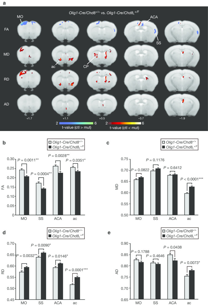
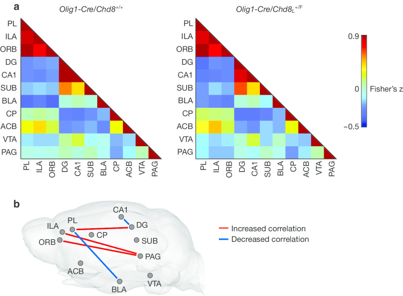
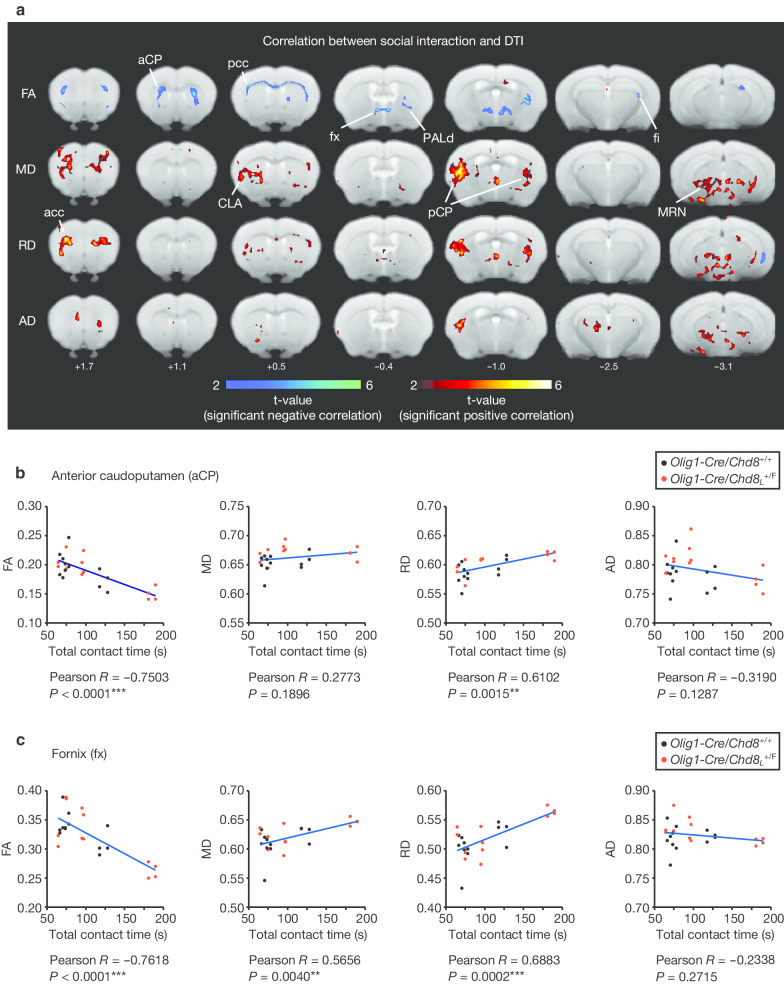
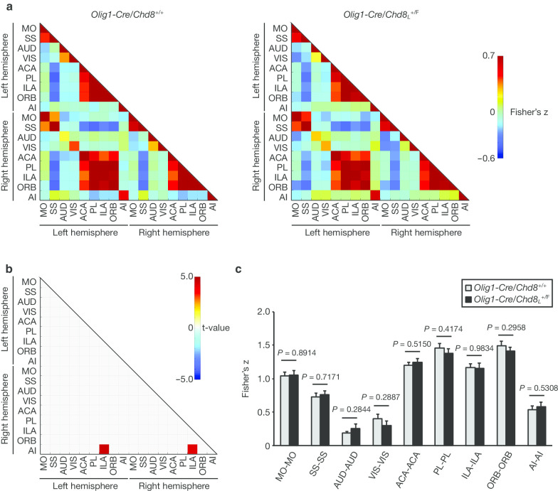
CHD8 encodes a chromatin-remodeling factor and is one of the most recurrently mutated genes in individuals with autism spectrum disorder (ASD). Although we have recently shown that mice heterozygous for Chd8 mutation manifest myelination defects and ASD-like behaviors, the detailed mechanisms underlying ASD pathogenesis have remained unclear. Here we performed diffusion tensor imaging (DTI) and resting-state functional magnetic resonance imaging (rsfMRI) in oligodendrocyte lineage-specific Chd8 heterozygous mutant mice. DTI revealed that ablation of Chd8 specifically in oligodendrocytes of mice was associated with microstructural changes of specific brain regions including the cortex and striatum. The extent of these changes in white matter including the corpus callosum and fornix was correlated with total contact time in the reciprocal social interaction test. Analysis with rsfMRI revealed changes in functional brain connectivity in the mutant mice, and the extent of such changes in the cortex, hippocampus, and amygdala was also correlated with the change in social interaction. Our results thus suggest that changes in brain microstructure and functional connectivity induced by oligodendrocyte dysfunction might underlie altered social interaction in mice with oligodendrocyte-specific CHD8 haploinsufficiency.
CHD8 编码一个染色质重塑因子,是自闭症谱系障碍(ASD)患者中最常发生突变的基因之一。虽然我们最近已经表明,杂合子突变的 Chd8 小鼠表现出髓鞘缺陷和 ASD 样行为,但 ASD 发病机制的详细机制仍不清楚。在这里,我们对少突胶质细胞谱系特异性 Chd8 杂合突变小鼠进行了弥散张量成像(DTI)和静息态功能磁共振成像(rsfMRI)。DTI 显示,Chd8 仅在小鼠的少突胶质细胞中缺失与包括皮质和纹状体在内的特定脑区的微观结构变化有关。这些变化的程度包括胼胝体和穹窿在内的白质与相互社交互动测试中的总接触时间相关。rsfMRI 的分析显示突变小鼠的功能脑连接发生变化,而皮质、海马体和杏仁核中这些变化的程度也与社交互动的变化相关。因此,我们的结果表明,少突胶质细胞功能障碍引起的脑微观结构和功能连接变化可能是 Chd8 杂合不足的少突胶质细胞特异性小鼠社交互动改变的基础。