Department of Neurosurgery, Maxine Dunitz Neurosurgical Research Institute, Cedars-Sinai Medical Center, 127 S. San Vicente Blvd., Los Angeles, CA, 90048, USA.
Department of Medicine, Cedars-Sinai Medical Center, Los Angeles, CA, 90048, USA.
Acta Neuropathol Commun. 2020 Nov 23;8(1):202. doi: 10.1186/s40478-020-01076-4.
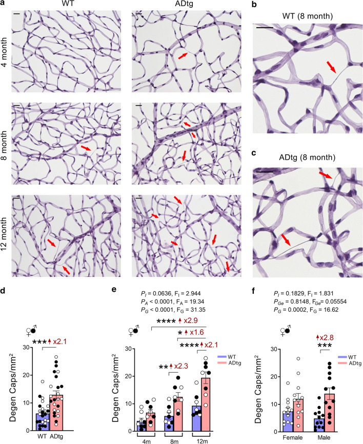
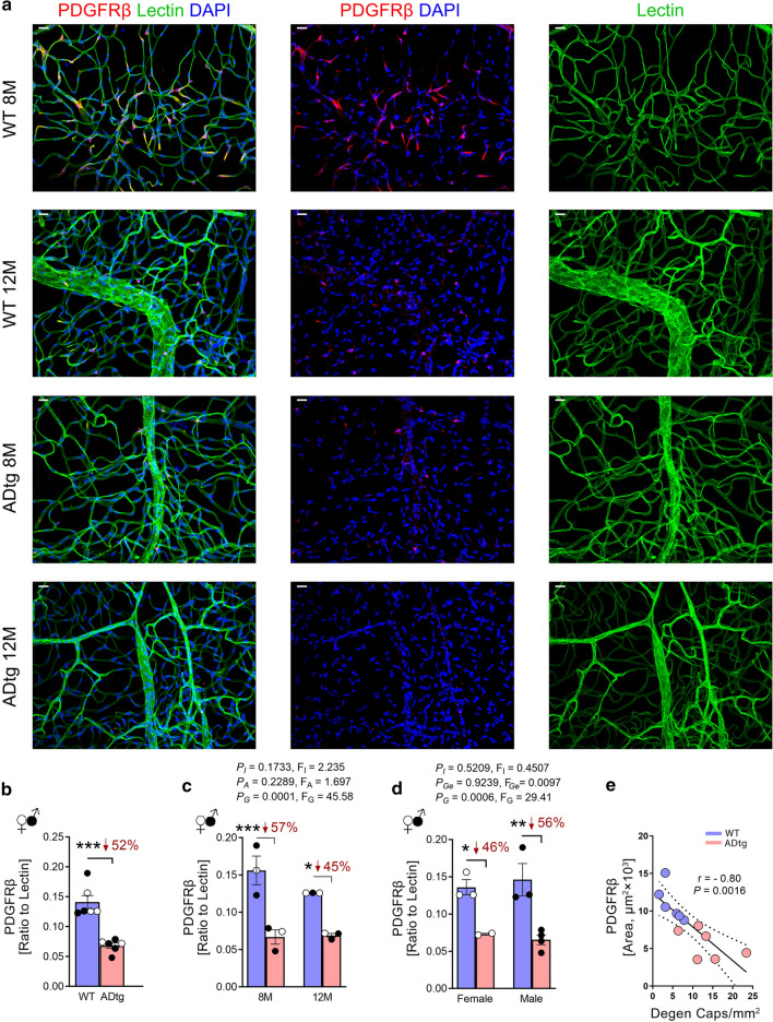
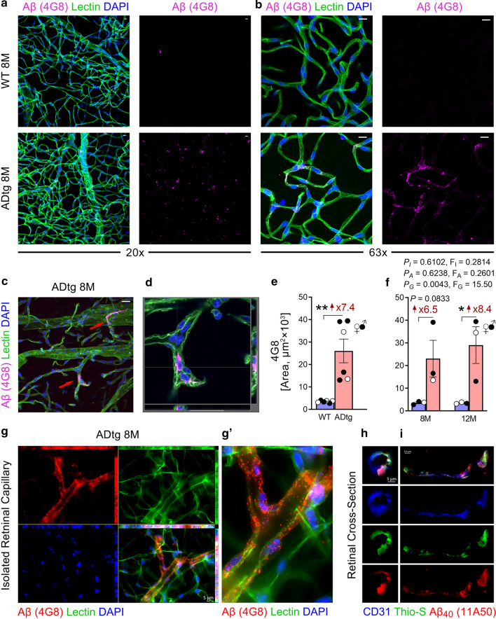
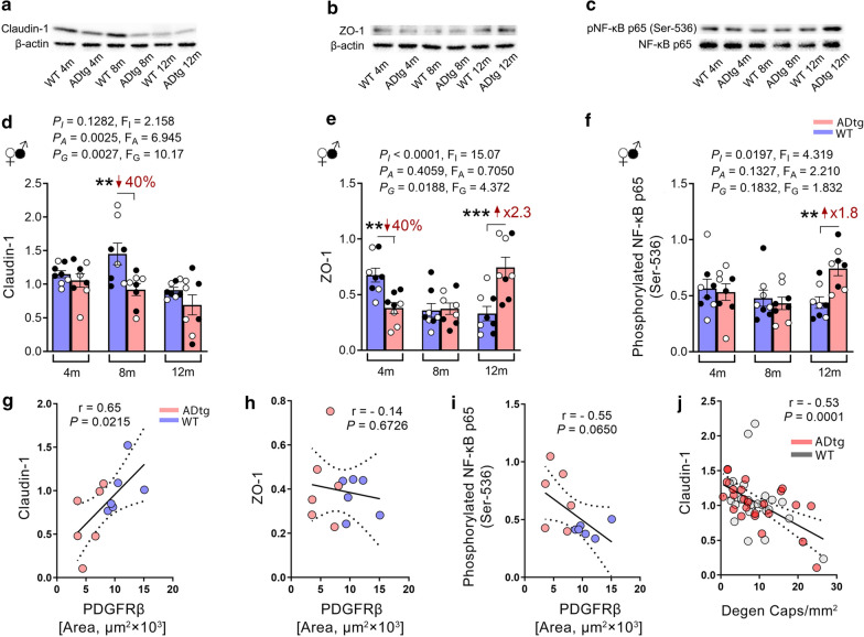
Extensive effort has been made studying retinal pathology in Alzheimer's disease (AD) to improve early noninvasive diagnosis and treatment. Particularly relevant are vascular changes, which appear prominent in early brain pathogenesis and could predict cognitive decline. Recently, we identified platelet-derived growth factor receptor beta (PDGFRβ) deficiency and pericyte loss associated with vascular Aβ deposition in the neurosensory retina of mild cognitively impaired (MCI) and AD patients. However, the pathological mechanisms of retinal vascular changes and their possible relationships with vascular amyloidosis, pericyte loss, and blood-retinal barrier (BRB) integrity remain unknown. Here, we evaluated the retinas of transgenic APP/PS1 mouse models of AD (ADtg mice) and wild-type mice at different ages for capillary degeneration, PDGFRβ expression, vascular amyloidosis, permeability and inner BRB tight-junction molecules. Using a retinal vascular isolation technique followed by periodic acid-Schiff or immunofluorescent staining, we discovered significant retinal capillary degeneration in ADtg mice compared to age- and sex-matched wild-type mice (P < 0.0001). This small vessel degeneration reached significance in 8-month-old mice (P = 0.0035), with males more susceptible than females. Degeneration of retinal capillaries also progressively increased with age in healthy mice (P = 0.0145); however, the phenomenon was significantly worse during AD-like progression (P = 0.0001). A substantial vascular PDGFRβ deficiency (~ 50% reduction, P = 0.0017) along with prominent vascular Aβ deposition was further detected in the retina of ADtg mice, which inversely correlated with the extent of degenerated capillaries (Pearson's r = - 0.8, P = 0.0016). Importantly, tight-junction alterations such as claudin-1 downregulation and increased BRB permeability, demonstrated in vivo by retinal fluorescein imaging and ex vivo following injection of FITC-dextran (2000 kD) and Texas Red-dextran (3 kD), were found in ADtg mice. Overall, the identification of age- and Alzheimer's-dependent retinal capillary degeneration and compromised BRB integrity starting at early disease stages in ADtg mice could contribute to the development of novel targets for AD diagnosis and therapy.
在阿尔茨海默病 (AD) 中,人们已经做出了大量努力来研究视网膜病理学,以改善早期的非侵入性诊断和治疗方法。特别相关的是血管变化,这些变化在早期的脑发病机制中表现得尤为明显,并且可以预测认知能力下降。最近,我们发现轻度认知障碍 (MCI) 和 AD 患者的神经感觉视网膜中与血管 Aβ 沉积相关的血小板衍生生长因子受体β (PDGFRβ) 缺乏和周细胞丧失。然而,视网膜血管变化的病理机制及其与血管淀粉样变性、周细胞丧失和血视网膜屏障 (BRB) 完整性的可能关系仍不清楚。在这里,我们评估了不同年龄的 APP/PS1 转基因 AD 小鼠模型 (ADtg 小鼠) 和野生型小鼠的视网膜,以研究毛细血管退化、PDGFRβ 表达、血管淀粉样变性、通透性和内 BRB 紧密连接分子。通过视网膜血管分离技术,随后进行过碘酸-Schiff 或免疫荧光染色,我们发现 ADtg 小鼠的视网膜毛细血管退化明显比年龄和性别匹配的野生型小鼠更严重 (P<0.0001)。这种小血管退化在 8 个月大的小鼠中具有统计学意义 (P=0.0035),且雄性比雌性更敏感。在健康小鼠中,视网膜毛细血管的退化也随年龄的增长而逐渐增加 (P=0.0145);然而,在类似 AD 的进展过程中,这种现象更为严重 (P=0.0001)。ADtg 小鼠的视网膜中还进一步检测到 PDGFRβ 明显缺乏 (~50%,P=0.0017) 和显著的血管 Aβ 沉积,这与退化的毛细血管程度呈负相关 (Pearson 相关系数 r=−0.8,P=0.0016)。重要的是,在 ADtg 小鼠中,通过视网膜荧光素成像体内检测到紧密连接改变,如 Claudin-1 下调和 BRB 通透性增加,并且在注射 FITC-葡聚糖 (2000 kD) 和 Texas Red-葡聚糖 (3 kD) 后体外也得到证实。总的来说,在 ADtg 小鼠中从疾病早期阶段开始出现年龄和阿尔茨海默病依赖性视网膜毛细血管退化和血视网膜屏障完整性受损,可能有助于开发用于 AD 诊断和治疗的新靶点。