• 文献检索
  • 文档翻译
  • 深度研究
  • 学术资讯
  • Suppr Zotero 插件Zotero 插件
  • 邀请有礼
  • 套餐&价格
  • 历史记录
应用&插件
Suppr Zotero 插件Zotero 插件浏览器插件Mac 客户端Windows 客户端微信小程序
定价
高级版会员购买积分包购买API积分包
服务
文献检索文档翻译深度研究API 文档MCP 服务
关于我们
关于 Suppr公司介绍联系我们用户协议隐私条款
关注我们

Suppr 超能文献

核心技术专利:CN118964589B侵权必究
粤ICP备2023148730 号-1Suppr @ 2026

文献检索

告别复杂PubMed语法,用中文像聊天一样搜索,搜遍4000万医学文献。AI智能推荐,让科研检索更轻松。

立即免费搜索

文件翻译

保留排版,准确专业,支持PDF/Word/PPT等文件格式,支持 12+语言互译。

免费翻译文档

深度研究

AI帮你快速写综述,25分钟生成高质量综述,智能提取关键信息,辅助科研写作。

立即免费体验

肝周间隙的磁共振成像

MR Imaging of the Perihepatic Space.

作者信息

Bonnin Angèle, Durot Carole, Djelouah Manel, Dohan Anthony, Arrivé Lionel, Rousset Pascal, Hoeffel Christine

机构信息

Department of Radiology, Centre Hospitalo-Universitaire de Reims, Reims, France.

Department of Abdominal and Interventional Radiology, Hôpital Cochin, Assistance Publique-Hôpitaux de Paris, APHP, Paris, France.

出版信息

Korean J Radiol. 2021 Apr;22(4):547-558. doi: 10.3348/kjr.2019.0774. Epub 2020 Nov 19.

DOI:10.3348/kjr.2019.0774
PMID:33236541
原文链接:https://pmc.ncbi.nlm.nih.gov/articles/PMC8005346/
Abstract

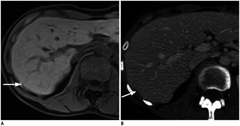
The perihepatic space is frequently involved in a spectrum of diseases, including intrahepatic lesions extending to the liver capsule and disease conditions involving adjacent organs extending to the perihepatic space or spreading thanks to the communication from intraperitoneal or extraperitoneal sites through the hepatic ligaments. Lesions resulting from the dissemination of peritoneal processes may also affect the perihepatic space. Here we discuss how to assess the perihepatic origin of a lesion and describe the magnetic resonance imaging (MRI) features of normal structures and fluids that may be abnormally located in the perihepatic space. We then review and illustrate the MRI findings present in cases of perihepatic infectious, tumor-related, and miscellaneous conditions. Finally, we highlight the value of MRI over computed tomography.

摘要

肝周间隙常累及一系列疾病,包括延伸至肝包膜的肝内病变以及累及相邻器官并延伸至肝周间隙的疾病状态,或因腹腔内或腹膜外部位通过肝韧带相通而蔓延至肝周间隙的疾病。腹膜病变播散所致的病变也可能影响肝周间隙。在此,我们讨论如何评估病变的肝周起源,并描述正常结构和液体在肝周间隙异常定位时的磁共振成像(MRI)特征。然后,我们回顾并举例说明肝周感染、肿瘤相关及其他情况的MRI表现。最后,我们强调MRI相对于计算机断层扫描的价值。

https://cdn.ncbi.nlm.nih.gov/pmc/blobs/d2f7/8005346/84d0f07a10d2/kjr-22-547-g014.jpg
https://cdn.ncbi.nlm.nih.gov/pmc/blobs/d2f7/8005346/b60350167be2/kjr-22-547-g001.jpg
https://cdn.ncbi.nlm.nih.gov/pmc/blobs/d2f7/8005346/a272c665673c/kjr-22-547-g002.jpg
https://cdn.ncbi.nlm.nih.gov/pmc/blobs/d2f7/8005346/49322786608d/kjr-22-547-g003.jpg
https://cdn.ncbi.nlm.nih.gov/pmc/blobs/d2f7/8005346/fc12859bf0a2/kjr-22-547-g004.jpg
https://cdn.ncbi.nlm.nih.gov/pmc/blobs/d2f7/8005346/47fd8f8ac85c/kjr-22-547-g005.jpg
https://cdn.ncbi.nlm.nih.gov/pmc/blobs/d2f7/8005346/bff4b3aacf4a/kjr-22-547-g006.jpg
https://cdn.ncbi.nlm.nih.gov/pmc/blobs/d2f7/8005346/109277d8e310/kjr-22-547-g007.jpg
https://cdn.ncbi.nlm.nih.gov/pmc/blobs/d2f7/8005346/a9d3f94726e2/kjr-22-547-g008.jpg
https://cdn.ncbi.nlm.nih.gov/pmc/blobs/d2f7/8005346/e9962fde581b/kjr-22-547-g009.jpg
https://cdn.ncbi.nlm.nih.gov/pmc/blobs/d2f7/8005346/f9f7256ceba5/kjr-22-547-g010.jpg
https://cdn.ncbi.nlm.nih.gov/pmc/blobs/d2f7/8005346/05c5bfc21c76/kjr-22-547-g011.jpg
https://cdn.ncbi.nlm.nih.gov/pmc/blobs/d2f7/8005346/87ad9bb4dd00/kjr-22-547-g012.jpg
https://cdn.ncbi.nlm.nih.gov/pmc/blobs/d2f7/8005346/e04aebbc5309/kjr-22-547-g013.jpg
https://cdn.ncbi.nlm.nih.gov/pmc/blobs/d2f7/8005346/84d0f07a10d2/kjr-22-547-g014.jpg

相似文献

1
MR Imaging of the Perihepatic Space.肝周间隙的磁共振成像
Korean J Radiol. 2021 Apr;22(4):547-558. doi: 10.3348/kjr.2019.0774. Epub 2020 Nov 19.
2
The perihepatic space: comprehensive anatomy and CT features of pathologic conditions.肝周间隙:病理状况的综合解剖及CT特征
Radiographics. 2007 Jan-Feb;27(1):129-43. doi: 10.1148/rg.271065050.
3
[CT & US findings could be useful for the diagnosis of Fitz-Hugh-Curtis syndrome].[CT和超声检查结果有助于诊断菲茨-休-柯蒂斯综合征]
Nihon Naika Gakkai Zasshi. 2015 Nov 10;104(11):2388-92. doi: 10.2169/naika.104.2388.
4
[Peritoneum and mesenterium. Radiological anatomy and extent of peritoneal diseases].[腹膜与肠系膜。腹膜疾病的放射解剖学及范围]
Radiologe. 2009 Jun;49(6):543-54; quiz 555-6. doi: 10.1007/s00117-008-1769-8.
5
Gadolinium-enhanced MR imaging of liver capsule and peritoneum.肝脏包膜和腹膜的钆增强磁共振成像。
Magn Reson Imaging Clin N Am. 2001 Nov;9(4):803-19, vii.
6
The peritoneum, mesenteries and omenta: normal anatomy and pathological processes.腹膜、肠系膜和网膜:正常解剖结构与病理过程
Eur Radiol. 1998;8(6):886-900. doi: 10.1007/s003300050485.
7
Fitz-Hugh-Curtis syndrome.菲茨-休-柯蒂斯综合征
Indian J Med Res. 2017 Jan;145(1):147. doi: 10.4103/ijmr.IJMR_1417_15.
8
Fitz-Hugh-Curtis syndrome.菲茨-休-柯蒂斯综合征
BMJ Case Rep. 2019 Feb 13;12(2):e229326. doi: 10.1136/bcr-2019-229326.
9
Fitz-Hugh-Curtis syndrome-like findings: chemical stimulation by the contrast agent.类菲茨-休-柯蒂斯综合征表现:造影剂的化学刺激。
Intern Med. 2013;52(22):2587-8. doi: 10.2169/internalmedicine.52.1173.
10
[Imaging of chronic pelvis pain].[慢性盆腔疼痛的影像学检查]
J Radiol. 2008 Jan;89(1 Pt 2):107-14. doi: 10.1016/s0221-0363(08)70381-0.

引用本文的文献

1
Cross-Sectional Imaging of Pelvic Inflammatory Disease: Diagnostic Pearls and Pitfalls on CT and MR.盆腔炎性疾病的横断面成像:CT和MR的诊断要点与陷阱
Diagnostics (Basel). 2025 Aug 10;15(16):2001. doi: 10.3390/diagnostics15162001.
2
Advances in Novel Diagnostic Techniques for Alveolar Echinococcosis.泡型包虫病新型诊断技术的进展
Diagnostics (Basel). 2025 Feb 27;15(5):585. doi: 10.3390/diagnostics15050585.
3
Migrating pseudolipoma of Glisson's capsule: A case report.肝门部假性脂肪瘤的迁移:一例报告。

本文引用的文献

1
Early cross-sectional imaging following open and laparoscopic cholecystectomy: a primer for radiologists.开腹和腹腔镜胆囊切除术后的早期横断面成像:放射科医生入门指南
Insights Imaging. 2018 Dec;9(6):925-941. doi: 10.1007/s13244-018-0663-9. Epub 2018 Nov 2.
2
MR imaging features of peritoneal alveolar echinococcosis.腹膜肺泡型棘球蚴病的磁共振成像特征
Diagn Interv Imaging. 2018 Jul-Aug;99(7-8):511-512. doi: 10.1016/j.diii.2018.02.009. Epub 2018 Mar 24.
3
Peritoneal and pleural fluids may appear hyperintense on hepatobiliary phase using hepatobiliary MR contrast agents.
Radiol Case Rep. 2024 Aug 29;19(11):5365-5369. doi: 10.1016/j.radcr.2024.08.028. eCollection 2024 Nov.
4
The role of multimodality imaging in the selection and management of patients treated with cytoreductive surgery and HIPEC.多模态影像学在细胞减灭术和 HIPEC 治疗患者的选择和管理中的作用。
Abdom Radiol (NY). 2024 Dec;49(12):4352-4364. doi: 10.1007/s00261-024-04441-2. Epub 2024 Jul 8.
5
Imaging Evaluation of Peritoneal Metastasis: Current and Promising Techniques.腹膜转移瘤的影像学评估:现有及有潜力的技术。
Korean J Radiol. 2024 Jan;25(1):86-102. doi: 10.3348/kjr.2023.0840.
6
Fitz-Hugh-Curtis Syndrome Presenting as Perihepatic and Subcapsular Enhancement on MRI.磁共振成像显示为肝周和包膜下强化的菲茨-休-柯蒂斯综合征
Case Rep Gastroenterol. 2022 Mar 31;16(1):235-239. doi: 10.1159/000523699. eCollection 2022 Jan-Apr.
使用肝胆磁共振对比剂,腹膜和胸腔积液在肝胆期可能表现为高信号。
Eur Radiol. 2018 Jul;28(7):3020-3031. doi: 10.1007/s00330-017-5261-6. Epub 2018 Jan 26.
4
Accuracy of apparent diffusion coefficient in differentiating pancreatic neuroendocrine tumour from intrapancreatic accessory spleen.表观扩散系数在鉴别胰腺神经内分泌肿瘤与胰内副脾中的准确性。
Eur Radiol. 2018 Apr;28(4):1560-1567. doi: 10.1007/s00330-017-5122-3. Epub 2017 Nov 13.
5
Evaluation of the peritoneal carcinomatosis index with CT and MRI.CT 和 MRI 评估腹膜癌转移指数。
Br J Surg. 2017 Aug;104(9):1244-1249. doi: 10.1002/bjs.10527. Epub 2017 Apr 4.
6
MRI in pelvic inflammatory disease: a pictorial review.MRI 在盆腔炎性疾病中的应用:影像学综述。
Abdom Radiol (NY). 2017 Mar;42(3):935-950. doi: 10.1007/s00261-016-1004-4.
7
Revised Atlanta Classification for Acute Pancreatitis: A Pictorial Essay.《急性胰腺炎的修订版亚特兰大分类:图文综述》
Radiographics. 2016 May-Jun;36(3):675-87. doi: 10.1148/rg.2016150097.
8
Radiology of epiploic appendages: acute appendagitis, post-infarcted appendages, and imaging natural history.脐旁附件的放射影像学:急性阑尾炎、梗死性附件炎和影像学自然病史。
Abdom Radiol (NY). 2016 Aug;41(8):1653-65. doi: 10.1007/s00261-016-0757-0.
9
Multimodality imaging in diagnosis and management of alveolar echinococcosis: an update.多模态影像学在细粒棘球蚴病的诊断和治疗中的应用:最新进展。
Diagn Interv Radiol. 2016 May-Jun;22(3):247-56. doi: 10.5152/dir.2015.15456.
10
MR diagnosis of diaphragmatic endometriosis.膈子宫内膜异位症的磁共振成像诊断
Eur Radiol. 2016 Nov;26(11):3968-3977. doi: 10.1007/s00330-016-4226-5. Epub 2016 Feb 12.