Department of Occupational and Environmental Health, School of Public Health, Capital Medical University, No. 10, Xitoutiao Youanmen Street, Beijing, 100069, China.
Beijing Key Laboratory of Environmental Toxicology, Capital Medical University, Beijing, 100069, China.
Stem Cell Res Ther. 2020 Nov 25;11(1):503. doi: 10.1186/s13287-020-02023-9.
Silicosis is an occupational respiratory disease caused by long-term excessive silica inhalation, which is most commonly encountered in industrial settings. Unfortunately, there is no effective therapy to delay and cure the progress of silicosis. In the recent years, stem cell therapy has emerged as an attractive tool against pulmonary fibrosis (PF) owing to its unique biological characteristics. However, the direct use of stem cells remains limitation by many risk factors for therapeutic purposes. The exclusive utility of exosomes secreted from stem cells, rather than cells, has been considered a promising alternative to overcome the limitations of cell-based therapy while maintaining its advantages.
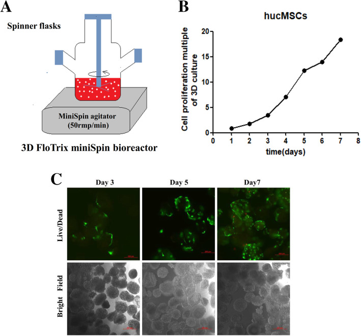
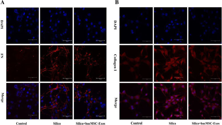
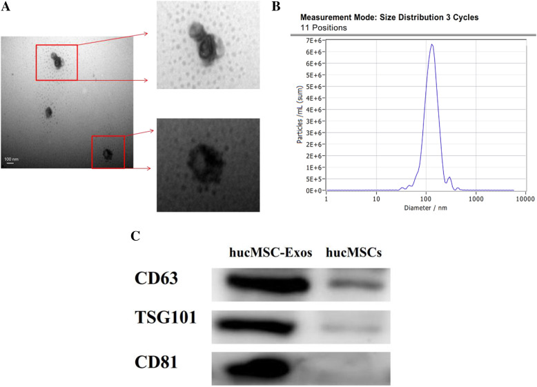
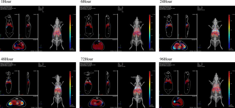
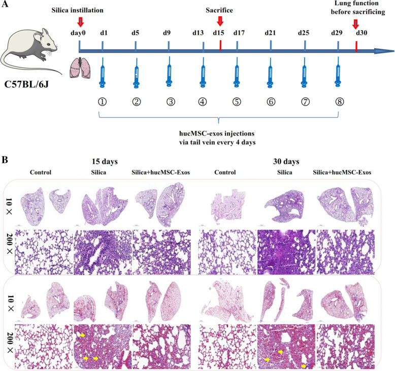
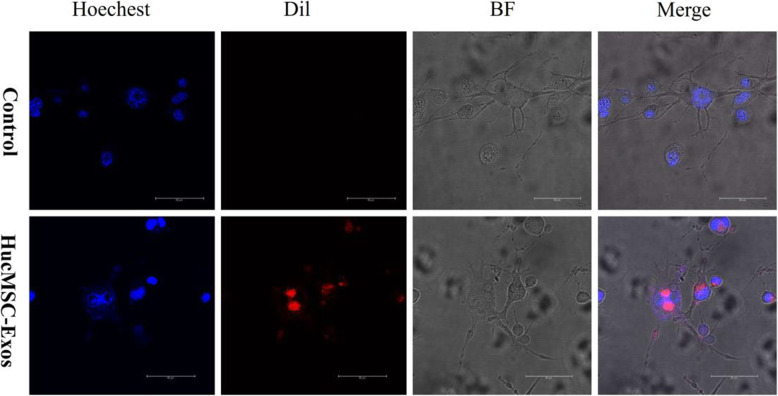
In this study, we first employed a three-dimensional (3D) dynamic system to culture human umbilical cord mesenchymal stem cell (hucMSC) spheroids in a microcarrier suspension to yield exosomes from serum-free media. Experimental silicosis was induced in C57BL/6J mice by intratracheal instillation of a silica suspension, with/without exosomes derived from hucMSC (hucMSC-Exos), injection via the tail vein afterwards. The results showed that the gene expression of collagen I (COL1A1) and fibronectin (FN) was upregulated in the silica group as compared to that in the control group; however, this change decreased with hucMSC-Exo treatment. The value of FEV0.1 decreased in the silica group as compared to that in the control group, and this change diminished with hucMSC-Exo treatment. These findings suggested that hucMSC-Exos could inhibit silica-induced PF and regulate pulmonary function. We also performed in vitro experiments to confirm these findings; the results revealed that hucMSC-Exos decreased collagen deposition in NIH-3T3 cells exposed to silica.
Taken together, these studies support a potential role for hucMSC-Exos in ameliorating pulmonary fibrosis and provide new evidence for improving clinical treatment induced by silica.
矽肺是一种由长期过量吸入二氧化硅引起的职业性呼吸道疾病,在工业环境中最为常见。不幸的是,目前尚无有效的治疗方法来延缓和阻止矽肺的进展。近年来,由于其独特的生物学特性,干细胞疗法作为一种治疗肺纤维化(PF)的有吸引力的工具而出现。然而,出于治疗目的,干细胞的直接应用仍受到许多风险因素的限制。人们认为,利用干细胞分泌的细胞外囊泡(exosomes)而不是细胞本身,是克服细胞治疗局限性的同时保持其优势的一种很有前途的替代方法。
在本研究中,我们首先采用三维(3D)动态系统在微载体悬浮液中培养人脐带间充质干细胞(hucMSC)球体,从无血清培养基中获得 exosomes。通过气管内滴注二氧化硅混悬液在 C57BL/6J 小鼠中诱导实验性矽肺,随后尾静脉注射 hucMSC 来源的 exosomes(hucMSC-Exos)。结果表明,与对照组相比,二氧化硅组胶原 I(COL1A1)和纤维连接蛋白(FN)的基因表达上调;然而,用 hucMSC-Exo 处理后,这种变化减少了。与对照组相比,二氧化硅组的 FEV0.1 值降低,用 hucMSC-Exo 处理后,这种变化减少了。这些发现表明 hucMSC-Exos 可以抑制二氧化硅诱导的 PF 并调节肺功能。我们还进行了体外实验来证实这些发现;结果表明,hucMSC-Exos 减少了暴露于二氧化硅的 NIH-3T3 细胞中的胶原蛋白沉积。
综上所述,这些研究支持 hucMSC-Exos 在改善肺纤维化中的潜在作用,并为改善二氧化硅诱导的临床治疗提供了新的证据。