Division of Imaging, Diagnostics and Software Reliability, Office of Science and Engineering Laboratories, Center for Devices and Radiological Health, Food and Drug Administration, Silver Spring, MD, 20993, USA.
Fischell Department of Bioengineering, University of Maryland, College Park, MD, 20742, USA.
Sci Rep. 2020 Nov 25;10(1):20505. doi: 10.1038/s41598-020-77554-5.
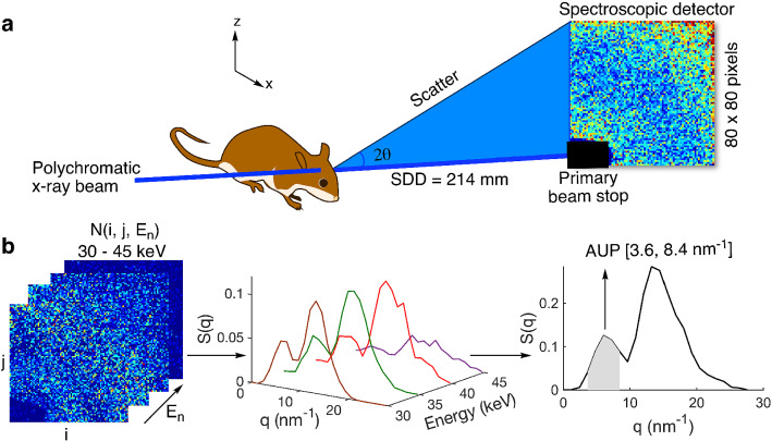
Amyloid plaque deposits in the brain are indicative of Alzheimer's and other diseases. Measurements of brain amyloid burden in small animals require laborious post-mortem histological analysis or resource-intensive, contrast-enhanced imaging techniques. We describe a label-free method based on spectral small-angle X-ray scattering with a polychromatic beam for in vivo estimation of brain amyloid burden. Our findings comparing 5XFAD versus wild-type mice correlate well with histology, showing promise for a fast and practical in vivo technique.
大脑中的淀粉样斑块沉积是阿尔茨海默病和其他疾病的标志物。小动物脑内淀粉样蛋白负担的测量需要费力的死后组织学分析或资源密集型的对比增强成像技术。我们描述了一种基于多色相干束的无标记光谱小角 X 射线散射方法,用于体内估计脑淀粉样蛋白负担。我们对 5XFAD 与野生型小鼠的比较研究结果与组织学相关性良好,为快速实用的体内技术提供了希望。