Division of Imaging, Diagnostics and Software Reliability, Office of Science and Engineering Laboratories, CDRH/FDA, Silver Spring, Maryland, United States of America.
PLoS One. 2020 Feb 11;15(2):e0228720. doi: 10.1371/journal.pone.0228720. eCollection 2020.
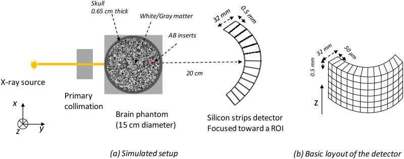
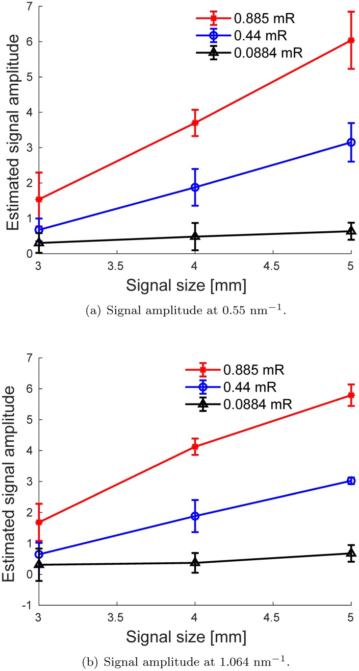
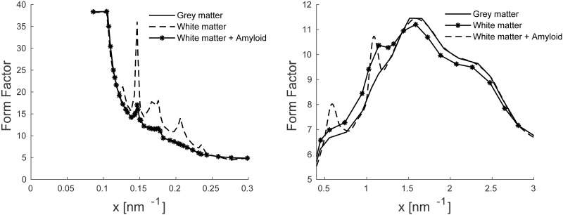
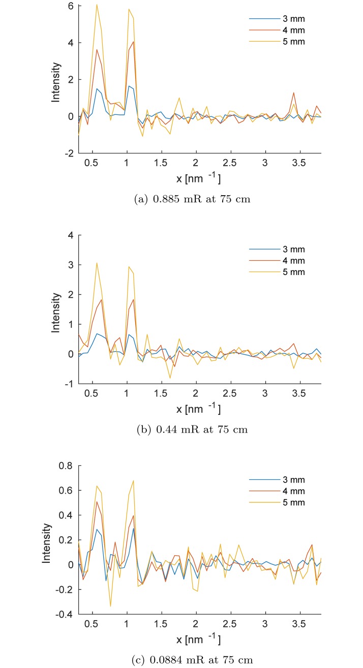
Brain aggregates of β amyloid (βA) protein plaques have been widely recognized as associated with many neurodegenerative diseases, and their identification can assist in the early diagnosis of Alzheimer's disease. We investigate the feasibility of using a spectral x-ray coherent scatter system with a silicon strip photon-counting detector for identifying brain βA protein plaques. This approach is based on differences in the structure of amyloid, white and grey matter in the brain. We simulated an energy- and angular-dispersive X-ray diffraction system with an x-ray pencil beam and Silicon strip sensor, energy-resolving detectors. The polychromatic beam is geometrically focused toward a region of interest in the brain. First, the open-source MC-GPU code for Monte Carlo transport was modified to accommodate the detector model. Second, brain phantoms with and without βA were simulated to assess the method and determine the radiation dose required to obtain acceptable statistical power. For βA targets of 3, 4 and 5 mm sizes in a 15-cm brain model, the required incident exposure was about 0.44 mR from a 60 kVp tungsten spectrum and 3.5 mm of added aluminum filtration. The results suggest that the proposed x-ray coherent scatter technique enables the use of high energy x-ray spectra and therefore has the potential to be used for accurate in vivo detection and quantification of βA in the brain within acceptable radiation dose levels.
脑内β淀粉样蛋白(βA)斑块的聚集物已被广泛认为与许多神经退行性疾病有关,其鉴定有助于阿尔茨海默病的早期诊断。我们研究了使用具有硅条光子计数探测器的光谱 X 射线相干散射系统来识别脑内βA 蛋白斑块的可行性。这种方法基于淀粉样蛋白、脑白质和灰质结构的差异。我们模拟了具有 X 射线铅笔束和硅条传感器的能量和角度色散 X 射线衍射系统,具有能量分辨探测器。多色光束在几何上聚焦到脑内的感兴趣区域。首先,修改了用于蒙特卡罗输运的开源 MC-GPU 代码以适应探测器模型。其次,模拟了有和没有βA 的脑模型,以评估该方法并确定获得可接受统计能力所需的辐射剂量。在 15cm 脑模型中,对于 3、4 和 5mm 大小的βA 靶标,从 60kVp 钨光谱和 3.5mm 附加铝过滤获得的所需入射曝光约为 0.44mR。结果表明,所提出的 X 射线相干散射技术能够使用高能 X 射线光谱,因此具有在可接受的辐射剂量水平内对脑内βA 进行准确体内检测和定量的潜力。