Department of Automatic Control and Robotics, AGH University of Science and Technology, 30-059 Kraków, Poland.
Cancer Biology Program, Department of Pathology, Stanford University School of Medicine, Stanford, CA 94305, USA.
Sensors (Basel). 2020 Nov 24;20(23):6713. doi: 10.3390/s20236713.
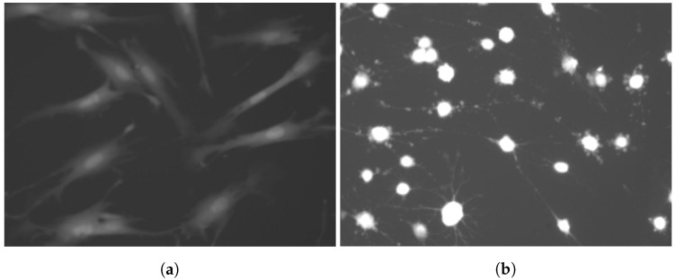
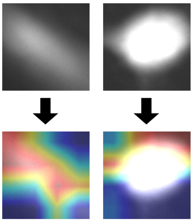
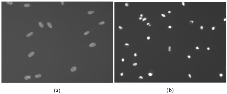
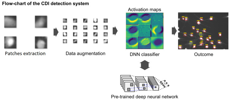
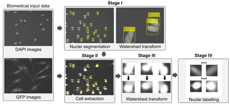
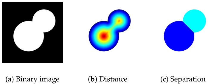
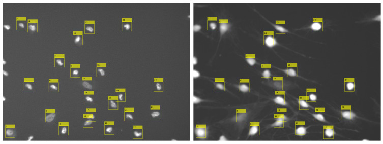
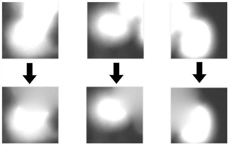
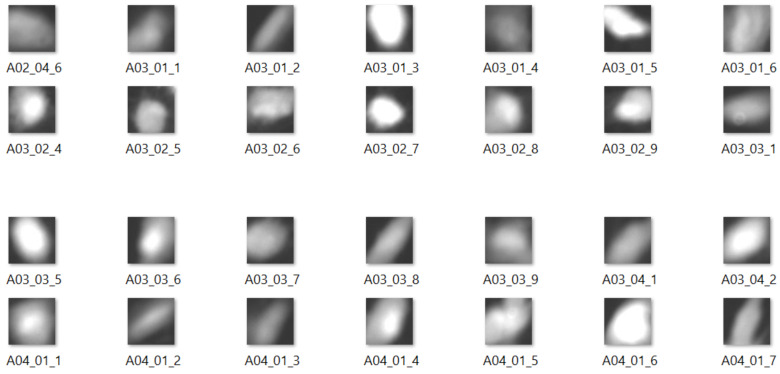
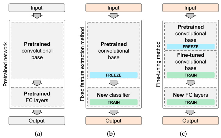
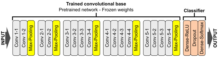
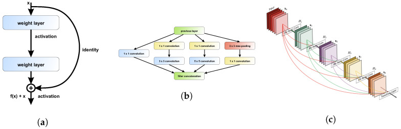
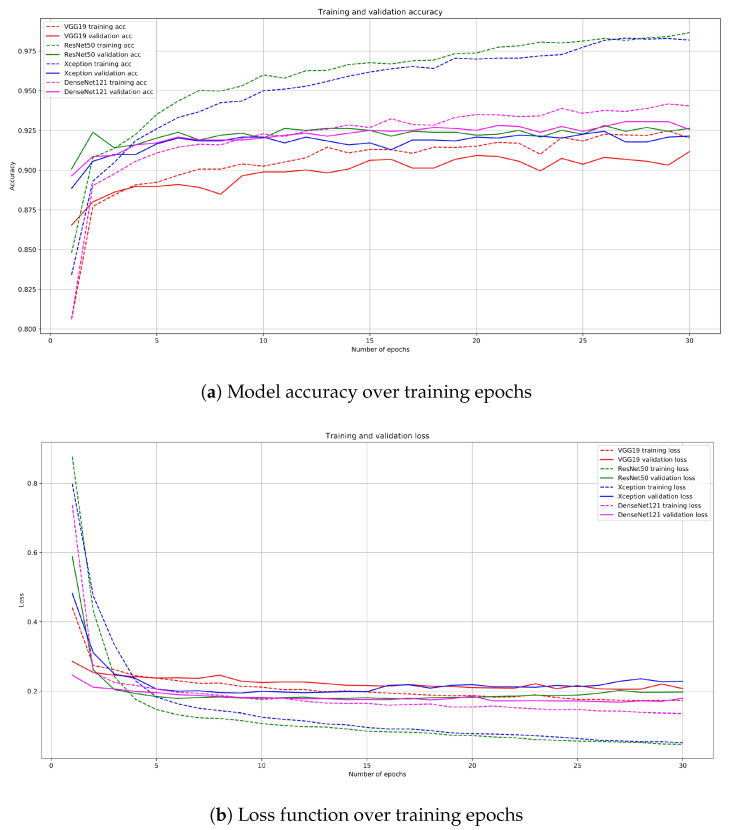
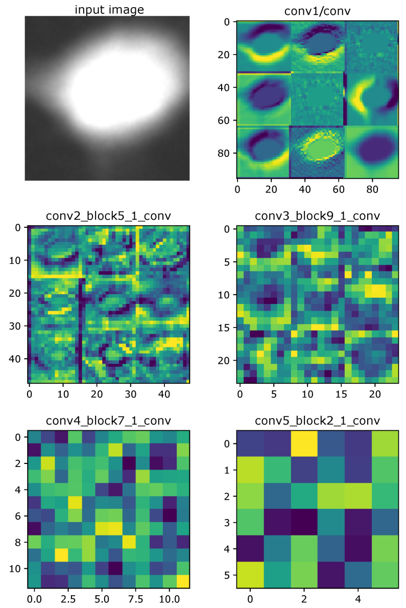
infection (CDI) is an enteric bacterial disease that is increasing in incidence worldwide. Symptoms of CDI range from mild diarrhea to severe life-threatening inflammation of the colon. While antibiotics are standard-of-care treatments for CDI, they are also the biggest risk factor for development of CDI and recurrence. Therefore, novel therapies that successfully treat CDI and protect against recurrence are an unmet clinical need. Screening for novel drug leads is often tested by manual image analysis. The process is slow, tedious and is subject to human error and bias. So far, little work has focused on computer-aided screening for drug leads based on fluorescence images. Here, we propose a novel method to identify characteristic morphological changes in human fibroblast cells exposed to toxins based on computer vision algorithms supported by deep learning methods. Classical image processing algorithms for the pre-processing stage are used together with an adjusted pre-trained deep convolutional neural network responsible for cell classification. In this study, we take advantage of transfer learning methodology by examining pre-trained VGG-19, ResNet50, Xception, and DenseNet121 convolutional neural network (CNN) models with adjusted, densely connected classifiers. We compare the obtained results with those of other machine learning algorithms and also visualize and interpret them. The proposed models have been evaluated on a dataset containing 369 images with 6112 cases. DenseNet121 achieved the highest results with a 93.5% accuracy, 92% sensitivity, and 95% specificity, respectively.
感染(CDI)是一种肠道细菌疾病,其发病率在全球范围内呈上升趋势。CDI 的症状范围从轻度腹泻到严重危及生命的结肠炎症。虽然抗生素是 CDI 的标准治疗方法,但它们也是 CDI 发展和复发的最大风险因素。因此,能够成功治疗 CDI 并预防复发的新型疗法是一种未满足的临床需求。新型药物先导物的筛选通常通过手动图像分析进行测试。这个过程缓慢、繁琐,并且容易受到人为错误和偏见的影响。到目前为止,很少有工作专注于基于荧光图像的计算机辅助药物先导物筛选。在这里,我们提出了一种新的方法,基于计算机视觉算法并结合深度学习方法,识别暴露于人源成纤维细胞毒素中的特征形态变化。用于预处理阶段的经典图像处理算法与负责细胞分类的调整后的预训练深度卷积神经网络一起使用。在这项研究中,我们通过检查经过调整的密集连接分类器的预训练 VGG-19、ResNet50、Xception 和 DenseNet121 卷积神经网络(CNN)模型,利用迁移学习方法。我们将获得的结果与其他机器学习算法进行比较,并且还对其进行可视化和解释。所提出的模型已经在包含 369 张图像和 6112 个病例的数据集上进行了评估。DenseNet121 分别实现了 93.5%的准确率、92%的灵敏度和 95%的特异性。