Perier-Metz Camille, Duda Georg N, Checa Sara
Julius Wolff Institute, Charité-Universitätsmedizin, Berlin, Germany.
MINES ParisTech - PSL Research University (Paris Sciences & Lettres), Paris, France.
Front Bioeng Biotechnol. 2020 Nov 11;8:585799. doi: 10.3389/fbioe.2020.585799. eCollection 2020.
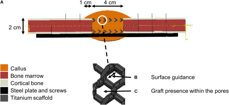
Large segmental bone defects represent a clinical challenge for which current treatment procedures have many drawbacks. 3D-printed scaffolds may help to support healing, but their design process relies mainly on trial and error due to a lack of understanding of which scaffold features support bone regeneration. The aim of this study was to investigate whether existing mechano-biological rules of bone regeneration can also explain scaffold-supported bone defect healing. In addition, we examined the distinct roles of bone grafting and scaffold structure on the regeneration process. To that end, scaffold-surface guided migration and tissue deposition as well as bone graft stimulatory effects were included in an model and predictions were compared to data. We found graft osteoconductive properties and scaffold-surface guided extracellular matrix deposition to be essential features driving bone defect filling in a 3D-printed honeycomb titanium structure. This knowledge paves the way for the design of more effective 3D scaffold structures and their pre-clinical optimization, prior to their application in scaffold-based bone defect regeneration.
大段骨缺损是一项临床挑战,当前的治疗方法存在诸多弊端。3D打印支架可能有助于促进愈合,但其设计过程主要依赖反复试验,因为缺乏对何种支架特征能支持骨再生的了解。本研究的目的是调查现有的骨再生机械生物学规则是否也能解释支架支持的骨缺损愈合情况。此外,我们研究了骨移植和支架结构在再生过程中的不同作用。为此,将支架表面引导的迁移和组织沉积以及骨移植刺激作用纳入一个模型,并将预测结果与实验数据进行比较。我们发现移植骨的骨传导特性和支架表面引导的细胞外基质沉积是驱动3D打印蜂窝状钛结构骨缺损填充的关键特征。这一知识为设计更有效的3D支架结构及其临床前优化铺平了道路,以便在基于支架的骨缺损再生中应用。