• 文献检索
  • 文档翻译
  • 深度研究
  • 学术资讯
  • Suppr Zotero 插件Zotero 插件
  • 邀请有礼
  • 套餐&价格
  • 历史记录
应用&插件
Suppr Zotero 插件Zotero 插件浏览器插件Mac 客户端Windows 客户端微信小程序
定价
高级版会员购买积分包购买API积分包
服务
文献检索文档翻译深度研究API 文档MCP 服务
关于我们
关于 Suppr公司介绍联系我们用户协议隐私条款
关注我们

Suppr 超能文献

核心技术专利:CN118964589B侵权必究
粤ICP备2023148730 号-1Suppr @ 2026

文献检索

告别复杂PubMed语法,用中文像聊天一样搜索,搜遍4000万医学文献。AI智能推荐,让科研检索更轻松。

立即免费搜索

文件翻译

保留排版,准确专业,支持PDF/Word/PPT等文件格式,支持 12+语言互译。

免费翻译文档

深度研究

AI帮你快速写综述,25分钟生成高质量综述,智能提取关键信息,辅助科研写作。

立即免费体验

多发性硬化中外层和中层黄斑层的功能评估

Functional Assessment of Outer and Middle Macular Layers in Multiple Sclerosis.

作者信息

Ziccardi Lucia, Barbano Lucilla, Boffa Laura, Albanese Maria, Nicoletti Carolina Gabri, Landi Doriana, Grzybowski Andrzej, Falsini Benedetto, Marfia Girolama Alessandra, Centonze Diego, Parisi Vincenzo

机构信息

IRCCS-Fondazione Bietti, Via Livenza 1, 00198 Rome, Italy.

Unit of Neurology, Fondazione Policlinico Tor Vergata, Via Oxford 81, 00133 Rome, Italy.

出版信息

J Clin Med. 2020 Nov 22;9(11):3766. doi: 10.3390/jcm9113766.

DOI:10.3390/jcm9113766
PMID:33266435
原文链接:https://pmc.ncbi.nlm.nih.gov/articles/PMC7700336/
Abstract

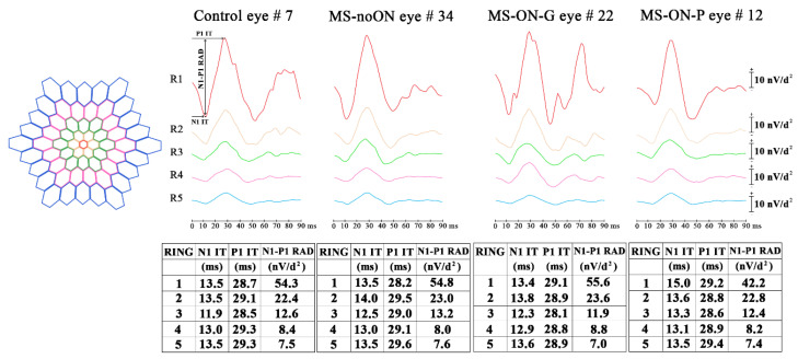
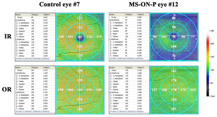
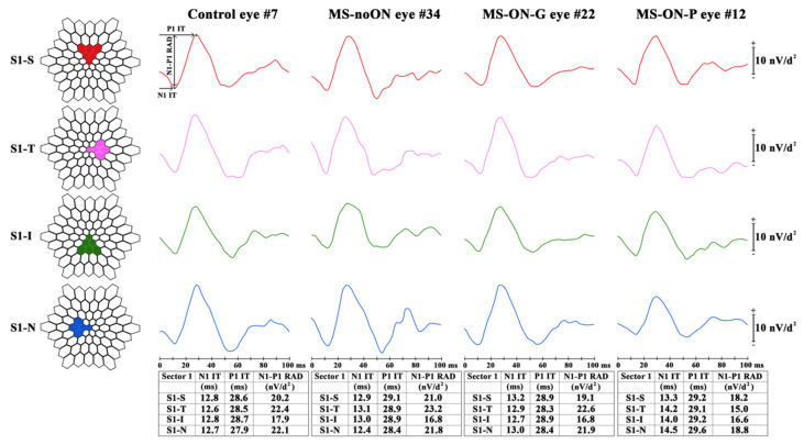
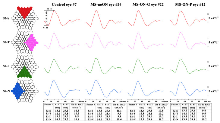
The involvement of macular preganglionic elements' function, during the neurodegenerative process of multiple sclerosis (MS), is controversial. In this case-control observational and retrospective study, we assessed multifocal electroretinogram (mfERG) responses from 41 healthy Controls, 41 relapsing-remitting MS patients without optic neuritis (ON) (MS-noON Group) and 47 MS patients with ON: 27 with full recovery of high-contrast best corrected visual acuity (BCVA) (MS-ON-G Group) and 20 with poor recovery (between 0.2 and 1 LogMAR) of BCVA, (MS-ON-P Group). In the latter Group, Sd-OCT macular volumes and thicknesses of whole and inner and outer retina were measured. MfERG N1 and P1 implicit times (ITs), and N1-P1 response amplitude densities (RADs), were measured from concentric rings (R) with increasing foveal eccentricity: 0-5° (R1), 5-10° (R2), 10-15° (R3), 15-20° (R4), 20-25° (R5), and from retinal sectors (superior, nasal, inferior and temporal) between 0-15° and 0-25°. In the MS-ON-P Group, mean mfERG RADs detected from R1 (0-5°) and from the central nasal sector (0-15°) were significantly reduced ( < 0.01) with respect to those of the Control, MS-noON and MS-ON-G Groups. No other significant differences between Groups for any mfERG parameters were found. All Sd-OCT measurements, apart from the inner retina macular volume in the central 1 mm, were significantly reduced in MS-ON-P patients compared to Controls. The functional impairment in the MS-ON-P Group was associated but not correlated with structural changes of the outer and inner retinal layers in corresponding retinal Areas and Sectors. Our results suggest that in MS, exclusively after ON with poor recovery of BCVA, the neurodegenerative process can induce dysfunctional mechanisms involving photoreceptors and bipolar cells of the fovea and of the more central nasal macular area.

摘要

在多发性硬化症(MS)的神经退行性变过程中,黄斑节前神经元功能的参与情况存在争议。在这项病例对照观察性回顾研究中,我们评估了41名健康对照者、41名无视神经炎(ON)的复发缓解型MS患者(MS-noON组)以及47名患有ON的MS患者的多焦视网膜电图(mfERG)反应:27名高对比度最佳矫正视力(BCVA)完全恢复的患者(MS-ON-G组)和20名BCVA恢复较差(0.2至1 LogMAR之间)的患者(MS-ON-P组)。在后者组中,测量了黄斑区的扫描光相干断层扫描(Sd-OCT)体积以及整个视网膜、视网膜内层和外层的厚度。从中心凹偏心度逐渐增加的同心环(R):0 - 5°(R1)、5 - 10°(R2)、10 - 15°(R3)、15 - 20°(R4)、20 - 25°(R5),以及0 - 15°和0 - 25°之间的视网膜象限(上、鼻、下和颞侧)测量mfERG的N1和P1隐时(ITs)以及N1 - P1反应振幅密度(RADs)。在MS-ON-P组中,从R1(0 - 5°)和中央鼻侧象限(0 - 15°)检测到的mfERG平均RADs相对于对照组、MS-noON组和MS-ON-G组显著降低(<0.01)。在任何mfERG参数方面,各组之间未发现其他显著差异。与对照组相比,MS-ON-P患者除中央1mm内视网膜黄斑体积外的所有Sd-OCT测量值均显著降低。MS-ON-P组的功能损害与相应视网膜区域和象限中外层和内层视网膜层的结构变化相关,但无相关性。我们的结果表明,在MS中,仅在ON后且BCVA恢复较差时,神经退行性变过程可诱导涉及中央凹和更中央鼻侧黄斑区的光感受器和双极细胞的功能障碍机制。

https://cdn.ncbi.nlm.nih.gov/pmc/blobs/8f69/7700336/badba4f46a60/jcm-09-03766-g004.jpg
https://cdn.ncbi.nlm.nih.gov/pmc/blobs/8f69/7700336/2ef3df36e1c9/jcm-09-03766-g001.jpg
https://cdn.ncbi.nlm.nih.gov/pmc/blobs/8f69/7700336/7ee41375d971/jcm-09-03766-g002.jpg
https://cdn.ncbi.nlm.nih.gov/pmc/blobs/8f69/7700336/6a3c6c7e6bb5/jcm-09-03766-g003.jpg
https://cdn.ncbi.nlm.nih.gov/pmc/blobs/8f69/7700336/badba4f46a60/jcm-09-03766-g004.jpg

相似文献

1
Functional Assessment of Outer and Middle Macular Layers in Multiple Sclerosis.多发性硬化中外层和中层黄斑层的功能评估
J Clin Med. 2020 Nov 22;9(11):3766. doi: 10.3390/jcm9113766.
2
Assessment of Macular Function by Multifocal Electroretinogram in Patients with Multiple Sclerosis Treated with Fingolimod.多发性硬化症患者接受芬戈莫德治疗后的多焦视网膜电图评估。
Adv Ther. 2021 Jul;38(7):3986-3996. doi: 10.1007/s12325-021-01728-4. Epub 2021 Jun 9.
3
Multifocal Electroretinogram Photopic Negative Response: A Reliable Paradigm to Detect Localized Retinal Ganglion Cells' Impairment in Retrobulbar Optic Neuritis Due to Multiple Sclerosis as a Model of Retinal Neurodegeneration.多焦视网膜电图明视负反应:一种可靠的检测范式,用于检测作为视网膜神经变性模型的多发性硬化所致球后视神经炎中局部视网膜神经节细胞的损伤。
Diagnostics (Basel). 2022 May 6;12(5):1156. doi: 10.3390/diagnostics12051156.
4
Multifocal electroretinogram in diabetic macular edema and its correlation with different optical coherence tomography features.多焦视网膜电图在糖尿病性黄斑水肿及其与不同光学相干断层扫描特征的相关性。
Int Ophthalmol. 2020 Mar;40(3):571-581. doi: 10.1007/s10792-019-01215-4. Epub 2019 Nov 11.
5
Retinal dysfunction characterizes subtypes of dominant optic atrophy.视网膜功能障碍是显性视神经萎缩亚型的特征。
Acta Ophthalmol. 2018 Mar;96(2):e156-e163. doi: 10.1111/aos.13557. Epub 2017 Sep 19.
6
Macular function in eyes with open-angle glaucoma evaluated by multifocal electroretinogram.多焦视网膜电图评估开角型青光眼患者的黄斑功能。
Invest Ophthalmol Vis Sci. 2012 Oct 9;53(11):6973-80. doi: 10.1167/iovs.12-10256.
7
Structure versus function: correlation between outer retinal and choroidal thicknesses measured by swept-source OCT with multifocal electroretinography and visual acuity.结构与功能:扫频源光学相干断层扫描测量的视网膜外层与脉络膜厚度之间的相关性与多焦视网膜电图及视力的关系
Int J Retina Vitreous. 2017 Aug 7;3:29. doi: 10.1186/s40942-017-0082-y. eCollection 2017.
8
Carotenoids and antioxidants in age-related maculopathy italian study: multifocal electroretinogram modifications after 1 year.年龄相关性黄斑病变中类胡萝卜素和抗氧化剂的意大利研究:1年后多焦视网膜电图的变化
Ophthalmology. 2008 Feb;115(2):324-333.e2. doi: 10.1016/j.ophtha.2007.05.029. Epub 2007 Aug 22.
9
Early and localized retinal dysfunction in patients with type 1 diabetes mellitus studied by multifocal electroretinogram.多焦视网膜电图研究 1 型糖尿病患者的早期和局部视网膜功能障碍。
Acta Diabetol. 2018 Nov;55(11):1191-1200. doi: 10.1007/s00592-018-1209-9. Epub 2018 Aug 28.
10
Morphological Outer Retina Findings in Multiple Sclerosis Patients With or Without Optic Neuritis.患有或未患有视神经炎的多发性硬化症患者的视网膜外层形态学表现
Front Neurol. 2020 Sep 15;11:858. doi: 10.3389/fneur.2020.00858. eCollection 2020.

引用本文的文献

1
Structural and Functional Assessment of the Macular Inner Retinal Layers in Multiple Sclerosis Eyes Without History of Optic Neuropathy.无视神经病变病史的多发性硬化症患者眼部黄斑区视网膜内层的结构和功能评估
J Clin Med. 2025 Aug 21;14(16):5919. doi: 10.3390/jcm14165919.
2
Evaluating the impact of anti-CGRP monoclonal antibodies on retinal features in migraine patients: a retrospective optical coherence tomography study.评估抗降钙素基因相关肽单克隆抗体对偏头痛患者视网膜特征的影响:一项回顾性光学相干断层扫描研究
Ther Adv Neurol Disord. 2025 Jun 24;18:17562864251347277. doi: 10.1177/17562864251347277. eCollection 2025.
3

本文引用的文献

1
Morphological Outer Retina Findings in Multiple Sclerosis Patients With or Without Optic Neuritis.患有或未患有视神经炎的多发性硬化症患者的视网膜外层形态学表现
Front Neurol. 2020 Sep 15;11:858. doi: 10.3389/fneur.2020.00858. eCollection 2020.
2
Commentary: Outer Retinal Dysfunction on Multifocal Electroretinography May Help Differentiating Multiple Sclerosis From Neuromyelitis Optica Spectrum Disorder.评论:多焦视网膜电图检测到的外层视网膜功能障碍可能有助于鉴别多发性硬化症与视神经脊髓炎谱系障碍。
Front Neurol. 2020 Apr 28;11:282. doi: 10.3389/fneur.2020.00282. eCollection 2020.
3
Decoding PERG: A neuro-ophthalmic retinal ganglion cell function review.
Retinal Function in Advanced Multiple Sclerosis.
晚期多发性硬化症的视网膜功能。
Invest Ophthalmol Vis Sci. 2024 Nov 4;65(13):2. doi: 10.1167/iovs.65.13.2.
4
Topographical Correlation between Structural and Functional Impairment of the Macular Inner Retinal Layers in Multiple Sclerosis Eyes with a History of Optic Neuropathy.多发性硬化症伴视神经病变史患者黄斑区视网膜内层结构与功能损害的地形学相关性
J Clin Med. 2023 Nov 19;12(22):7175. doi: 10.3390/jcm12227175.
5
Multifocal Electroretinogram Photopic Negative Response: A Reliable Paradigm to Detect Localized Retinal Ganglion Cells' Impairment in Retrobulbar Optic Neuritis Due to Multiple Sclerosis as a Model of Retinal Neurodegeneration.多焦视网膜电图明视负反应:一种可靠的检测范式,用于检测作为视网膜神经变性模型的多发性硬化所致球后视神经炎中局部视网膜神经节细胞的损伤。
Diagnostics (Basel). 2022 May 6;12(5):1156. doi: 10.3390/diagnostics12051156.
6
Lyso-Lipid-Induced Oligodendrocyte Maturation Underlies Restoration of Optic Nerve Function.溶血磷脂诱导少突胶质细胞成熟是视神经功能恢复的基础。
eNeuro. 2022 Jan 26;9(1). doi: 10.1523/ENEURO.0429-21.2022. Print 2022 Jan-Feb.
7
Macular Morpho-Functional and Visual Pathways Functional Assessment in Patients with Spinocerebellar Type 1 Ataxia with or without Neurological Signs.伴或不伴神经体征的脊髓小脑性共济失调1型患者的黄斑形态功能及视觉通路功能评估
J Clin Med. 2021 Nov 12;10(22):5271. doi: 10.3390/jcm10225271.
8
OCT Variability Prevents Their Use as Robust Biomarkers in Multiple Sclerosis.光学相干断层扫描(OCT)的变异性使其无法作为多发性硬化症中可靠的生物标志物。
Clin Ophthalmol. 2021 May 14;15:2025-2036. doi: 10.2147/OPTH.S309703. eCollection 2021.
解读图形视网膜电图:神经眼科视网膜神经节细胞功能综述
Curr Ophthalmol Rep. 2019 Mar;7(1):51-58. doi: 10.1007/s40135-019-00199-9. Epub 2019 Feb 13.
4
Diagnostic ability of multifocal electroretinogram in early multiple sclerosis using a new signal analysis method.多焦视网膜电图在使用新信号分析方法诊断早期多发性硬化症中的诊断能力。
PLoS One. 2019 Nov 8;14(11):e0224500. doi: 10.1371/journal.pone.0224500. eCollection 2019.
5
Outer Retinal Dysfunction on Multifocal Electroretinography May Help Differentiating Multiple Sclerosis From Neuromyelitis Optica Spectrum Disorder.多焦视网膜电图显示的外层视网膜功能障碍可能有助于鉴别多发性硬化症和视神经脊髓炎谱系障碍。
Front Neurol. 2019 Aug 27;10:928. doi: 10.3389/fneur.2019.00928. eCollection 2019.
6
Functional Changes of Retinal Ganglion Cells and Visual Pathways in Patients with Chronic Leber's Hereditary Optic Neuropathy during One Year of Follow-up.慢性莱伯遗传性视神经病变患者在一年随访期内视网膜神经节细胞和视觉通路的功能变化。
Ophthalmology. 2019 Jul;126(7):1033-1044. doi: 10.1016/j.ophtha.2019.02.018. Epub 2019 Feb 26.
7
Functional architecture of the foveola revealed in the living primate.活灵长类动物中揭示的中央小凹的功能结构。
PLoS One. 2018 Nov 28;13(11):e0207102. doi: 10.1371/journal.pone.0207102. eCollection 2018.
8
Early auto-immune targeting of photoreceptor ribbon synapses in mouse models of multiple sclerosis.多发性硬化症小鼠模型中光感受器带突触的早期自身免疫靶向。
EMBO Mol Med. 2018 Nov;10(11). doi: 10.15252/emmm.201808926.
9
Early and localized retinal dysfunction in patients with type 1 diabetes mellitus studied by multifocal electroretinogram.多焦视网膜电图研究 1 型糖尿病患者的早期和局部视网膜功能障碍。
Acta Diabetol. 2018 Nov;55(11):1191-1200. doi: 10.1007/s00592-018-1209-9. Epub 2018 Aug 28.
10
ISCEV guide to visual electrodiagnostic procedures.国际临床视觉电生理学会视觉电诊断程序指南
Doc Ophthalmol. 2018 Feb;136(1):1-26. doi: 10.1007/s10633-017-9621-y. Epub 2018 Feb 3.