• 文献检索
  • 文档翻译
  • 深度研究
  • 学术资讯
  • Suppr Zotero 插件Zotero 插件
  • 邀请有礼
  • 套餐&价格
  • 历史记录
应用&插件
Suppr Zotero 插件Zotero 插件浏览器插件Mac 客户端Windows 客户端微信小程序
定价
高级版会员购买积分包购买API积分包
服务
文献检索文档翻译深度研究API 文档MCP 服务
关于我们
关于 Suppr公司介绍联系我们用户协议隐私条款
关注我们

Suppr 超能文献

核心技术专利:CN118964589B侵权必究
粤ICP备2023148730 号-1Suppr @ 2026

文献检索

告别复杂PubMed语法,用中文像聊天一样搜索,搜遍4000万医学文献。AI智能推荐,让科研检索更轻松。

立即免费搜索

文件翻译

保留排版,准确专业,支持PDF/Word/PPT等文件格式,支持 12+语言互译。

免费翻译文档

深度研究

AI帮你快速写综述,25分钟生成高质量综述,智能提取关键信息,辅助科研写作。

立即免费体验

多焦视网膜电图在使用新信号分析方法诊断早期多发性硬化症中的诊断能力。

Diagnostic ability of multifocal electroretinogram in early multiple sclerosis using a new signal analysis method.

机构信息

Biomedical Engineering Group, Electronics Department, Universidad de Alcalá, Alcalá de Henares, Madrid, Spain.

RETICS: Thematic Networks for Co-operative Research in Health for Ocular Diseases, Madrid, Spain.

出版信息

PLoS One. 2019 Nov 8;14(11):e0224500. doi: 10.1371/journal.pone.0224500. eCollection 2019.

DOI:10.1371/journal.pone.0224500
PMID:31703082
原文链接:https://pmc.ncbi.nlm.nih.gov/articles/PMC6839873/
Abstract

PURPOSE

To determine if a novel analysis method will increase the diagnostic value of the multifocal electroretinogram (mfERG) in diagnosing early-stage multiple sclerosis (MS).

METHODS

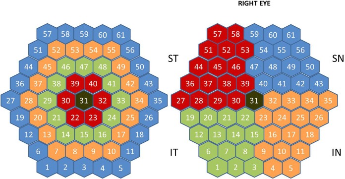
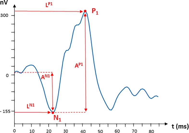
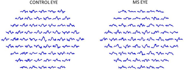
We studied the mfERG signals of OD (Oculus Dexter) eyes of fifteen patients diagnosed with early-stage MS (in all cases < 12 months) and without a history of optic neuritis (ON) (F:M = 11:4), and those of six controls (F:M = 3:3). We obtained values of amplitude and latency of N1 and P1 waves, and a method to assess normalized root-mean-square error (FNRMSE) between model signals and mfERG recordings was used. Responses of each eye were analysed at a global level, and by rings, quadrants and hemispheres. AUC (area under the ROC curve) is used as discriminant factor.

RESULTS

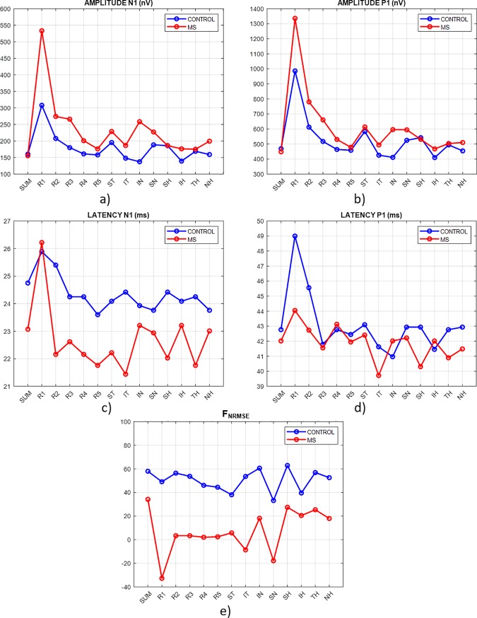
The standard method of analysis obtains further discrimination between controls and MS in ring R3 (AUC = 0.82), analysing N1 waves amplitudes. In all of the retina analysis regions, FNRMSE value shows a greater discriminating power than the standard method. The highest AUC value (AUC = 0.91) was in the superior temporal quadrant.

CONCLUSION

By analysing mfERG recordings and contrasting them with those of healthy controls it is possible to detect early-stage MS in patients without a previous history of ON.

摘要

目的

确定一种新的分析方法是否会提高多焦视网膜电图(mfERG)在诊断早期多发性硬化症(MS)中的诊断价值。

方法

我们研究了 15 名被诊断为早期 MS(所有病例均<12 个月)且无视神经炎(ON)病史的患者的右眼 mfERG 信号(F:M=11:4),以及 6 名对照者的右眼 mfERG 信号(F:M=3:3)。我们获得了 N1 和 P1 波的振幅和潜伏期值,并使用了一种评估模型信号与 mfERG 记录之间归一化均方根误差(FNRMSE)的方法。对每只眼睛进行了全局水平和环、象限和半球的分析。AUC(ROC 曲线下面积)用作判别因子。

结果

标准分析方法在分析 N1 波振幅的 R3 环(AUC=0.82)中进一步区分了对照组和 MS 患者。在所有视网膜分析区域中,FNRMSE 值的判别能力均强于标准方法。最高 AUC 值(AUC=0.91)位于颞上象限。

结论

通过分析 mfERG 记录并将其与健康对照者的记录进行对比,可以检测出无先前 ON 病史的早期 MS 患者。

https://cdn.ncbi.nlm.nih.gov/pmc/blobs/cf4a/6839873/252caa5d9270/pone.0224500.g005.jpg
https://cdn.ncbi.nlm.nih.gov/pmc/blobs/cf4a/6839873/78d4f88ca608/pone.0224500.g001.jpg
https://cdn.ncbi.nlm.nih.gov/pmc/blobs/cf4a/6839873/209d001bb27b/pone.0224500.g002.jpg
https://cdn.ncbi.nlm.nih.gov/pmc/blobs/cf4a/6839873/783a29c8f7f3/pone.0224500.g003.jpg
https://cdn.ncbi.nlm.nih.gov/pmc/blobs/cf4a/6839873/63e33f97ce12/pone.0224500.g004.jpg
https://cdn.ncbi.nlm.nih.gov/pmc/blobs/cf4a/6839873/252caa5d9270/pone.0224500.g005.jpg

相似文献

1
Diagnostic ability of multifocal electroretinogram in early multiple sclerosis using a new signal analysis method.多焦视网膜电图在使用新信号分析方法诊断早期多发性硬化症中的诊断能力。
PLoS One. 2019 Nov 8;14(11):e0224500. doi: 10.1371/journal.pone.0224500. eCollection 2019.
2
Identification of clusters in multifocal electrophysiology recordings to maximize discriminant capacity (patients vs. control subjects).在多焦点电生理记录中识别聚类以最大化判别能力(患者与对照受试者)。
Doc Ophthalmol. 2020 Feb;140(1):43-53. doi: 10.1007/s10633-019-09720-8. Epub 2019 Sep 19.
3
Empirical Mode Decomposition-Based Filter Applied to Multifocal Electroretinograms in Multiple Sclerosis Diagnosis.基于经验模态分解的滤波器在多发性硬化症诊断中的多焦视网膜电图应用。
Sensors (Basel). 2019 Dec 18;20(1):7. doi: 10.3390/s20010007.
4
Is multifocal electroretinography an early predictor of glaucoma?多焦视网膜电图是青光眼的早期预测指标吗?
Doc Ophthalmol. 2016 Feb;132(1):27-37. doi: 10.1007/s10633-016-9524-3. Epub 2016 Jan 20.
5
The 2-global flash mfERG in glaucoma: attempting to increase sensitivity by reducing the focal flash luminance and changing filter settings.青光眼患者的双眼全视野闪光多焦视网膜电图:试图通过降低局部闪光亮度和改变滤波设置来提高敏感性。
Doc Ophthalmol. 2013 Feb;126(1):57-67. doi: 10.1007/s10633-012-9360-z. Epub 2012 Nov 20.
6
Assessment of Macular Function by Multifocal Electroretinogram in Patients with Multiple Sclerosis Treated with Fingolimod.多发性硬化症患者接受芬戈莫德治疗后的多焦视网膜电图评估。
Adv Ther. 2021 Jul;38(7):3986-3996. doi: 10.1007/s12325-021-01728-4. Epub 2021 Jun 9.
7
Effect of axial length on full-field and multifocal electroretinograms.眼轴长度对全视野和多焦视网膜电图的影响。
Clin Exp Optom. 2017 Nov;100(6):668-675. doi: 10.1111/cxo.12529. Epub 2017 Mar 7.
8
Empirical mode decomposition processing to improve multifocal-visual-evoked-potential signal analysis in multiple sclerosis.采用经验模态分解处理改善多发性硬化症多焦点视觉诱发电位信号分析。
PLoS One. 2018 Apr 20;13(4):e0194964. doi: 10.1371/journal.pone.0194964. eCollection 2018.
9
Multifocal electroretinogram responses in Nepalese diabetic patients without retinopathy.尼泊尔无视网膜病变糖尿病患者的多焦视网膜电图反应
Doc Ophthalmol. 2014 Aug;129(1):39-46. doi: 10.1007/s10633-014-9447-9. Epub 2014 Jun 5.
10
Central retinal function as measured by the multifocal electroretinogram and flicker perimetry in early age-related macular degeneration.早期年龄相关性黄斑变性的多焦视网膜电图和闪烁视敏度测量的中心视网膜功能。
Invest Ophthalmol Vis Sci. 2011 Nov 29;52(12):9267-74. doi: 10.1167/iovs.11-8517.

引用本文的文献

1
Early detection and staging of retinitis pigmentosa using multifocal electroretinogram parameters and machine learning algorithms.使用多焦视网膜电图参数和机器学习算法对视网膜色素变性进行早期检测和分期。
Phys Eng Sci Med. 2025 Jun 16. doi: 10.1007/s13246-025-01577-3.
2
Retinal Function in Advanced Multiple Sclerosis.晚期多发性硬化症的视网膜功能。
Invest Ophthalmol Vis Sci. 2024 Nov 4;65(13):2. doi: 10.1167/iovs.65.13.2.
3
Lyso-Lipid-Induced Oligodendrocyte Maturation Underlies Restoration of Optic Nerve Function.溶血磷脂诱导少突胶质细胞成熟是视神经功能恢复的基础。

本文引用的文献

1
ISCEV guide to visual electrodiagnostic procedures.国际临床视觉电生理学会视觉电诊断程序指南
Doc Ophthalmol. 2018 Feb;136(1):1-26. doi: 10.1007/s10633-017-9621-y. Epub 2018 Feb 3.
2
Outer Retinal Dysfunction in the Absence of Structural Abnormalities in Multiple Sclerosis.多发性硬化症中无结构异常的视网膜外层功能障碍。
Invest Ophthalmol Vis Sci. 2018 Jan 1;59(1):549-560. doi: 10.1167/iovs.17-22821.
3
Diagnosis of multiple sclerosis: 2017 revisions of the McDonald criteria.多发性硬化症的诊断:2017 年麦当劳标准修订版。
eNeuro. 2022 Jan 26;9(1). doi: 10.1523/ENEURO.0429-21.2022. Print 2022 Jan-Feb.
4
Macular Morpho-Functional and Visual Pathways Functional Assessment in Patients with Spinocerebellar Type 1 Ataxia with or without Neurological Signs.伴或不伴神经体征的脊髓小脑性共济失调1型患者的黄斑形态功能及视觉通路功能评估
J Clin Med. 2021 Nov 12;10(22):5271. doi: 10.3390/jcm10225271.
5
Functional Assessment of Outer and Middle Macular Layers in Multiple Sclerosis.多发性硬化中外层和中层黄斑层的功能评估
J Clin Med. 2020 Nov 22;9(11):3766. doi: 10.3390/jcm9113766.
Lancet Neurol. 2018 Feb;17(2):162-173. doi: 10.1016/S1474-4422(17)30470-2. Epub 2017 Dec 21.
4
Retinal and Choroidal Changes in Patients with Parkinson's Disease Detected by Swept-Source Optical Coherence Tomography.扫频光学相干断层扫描检测帕金森病患者的视网膜和脉络膜变化
Curr Eye Res. 2018 Jan;43(1):109-115. doi: 10.1080/02713683.2017.1370116. Epub 2017 Nov 7.
5
OCT Glaucoma Staging System: a new method for retinal nerve fiber layer damage classification using spectral-domain OCT.光学相干断层扫描(OCT)青光眼分期系统:一种使用频域 OCT 对视网膜神经纤维层损伤进行分类的新方法。
Eye (Lond). 2018 Jan;32(1):113-119. doi: 10.1038/eye.2017.159. Epub 2017 Aug 4.
6
MRI in the assessment and monitoring of multiple sclerosis: an update on best practice.磁共振成像在多发性硬化症评估与监测中的应用:最佳实践的最新进展
Ther Adv Neurol Disord. 2017 Jun;10(6):247-261. doi: 10.1177/1756285617708911. Epub 2017 May 12.
7
A new role for evoked potentials in MS? Repurposing evoked potentials as biomarkers for clinical trials in MS.诱发电位在多发性硬化症中的新作用?将诱发电位重新用作多发性硬化症临床试验的生物标志物。
Mult Scler. 2017 Sep;23(10):1309-1319. doi: 10.1177/1352458517707265. Epub 2017 May 8.
8
Retinal and Optic Nerve Degeneration in Patients with Multiple Sclerosis Followed up for 5 Years.多发性硬化症患者随访 5 年的视网膜和视神经变性。
Ophthalmology. 2017 May;124(5):688-696. doi: 10.1016/j.ophtha.2017.01.005. Epub 2017 Feb 7.
9
Multimodal evoked potentials for functional quantification and prognosis in multiple sclerosis.用于多发性硬化症功能量化和预后评估的多模态诱发电位
BMC Neurol. 2016 Jun 1;16:83. doi: 10.1186/s12883-016-0608-1.
10
M-sequences in ophthalmic electrophysiology.眼科电生理学中的M序列
J Vis. 2016;16(1):15. doi: 10.1167/16.1.15.